The radius and ulna, the two bones of the forearm, have distinct anatomical features that necessitate different approaches in fracture management. While the ulna is relatively straight, the radius boasts a characteristic bow crucial for forearm rotation. This article explores the implications of this anatomical difference, focusing on the fit of pre-contoured plates used in radial shaft fracture fixation.
Understanding the Anatomical Differences: Radius vs. Ulna
The radius and ulna articulate at both the proximal and distal radioulnar joints, allowing for the complex rotational movements of the forearm. The radius’s distinctive bow, a crucial element of this biomechanics, requires careful consideration during fracture fixation. Failure to accurately restore this bow can significantly impact forearm function, leading to reduced range of motion and compromised joint mechanics. [1-5] Consequently, radial fractures often necessitate the use of contoured plates, meticulously shaped to mirror the bone’s natural curvature in three dimensions: sagittal, coronal, and potentially axial. [6-8]
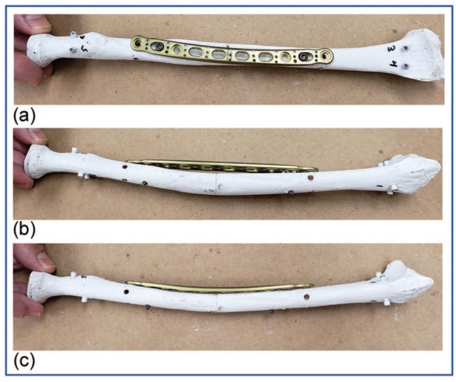
Figure 1: A plate secured to the radius with measuring holes for assessing bone-plate gap.
Comparing Pre-Contoured and Surgeon-Contoured Plates: A Cadaveric Study
A study investigated the fit of pre-contoured radial shaft plates compared to surgeon-contoured plates using cadaveric radii. Pre-contoured volar and dorsolateral plates were compared to straight plates manually contoured by a surgeon. Measurements were taken to quantify the gap between the plate and the bone surface, providing insights into the accuracy of the pre-contoured plates.
Materials and Methods: Assessing Plate Fit
Six cadaveric radii were used to assess the fit of three different plate types: pre-contoured volar, pre-contoured dorsolateral, and straight plates contoured by a hand surgeon. The plates were secured to the radii, and a digital depth gauge was used to measure the gap between the plate and the bone at pre-drilled holes. Measurements were taken for each plate type, including pre-contoured plates before and after additional surgeon contouring. Surgeon contouring time was also recorded.
Results: Dorsal vs. Volar Plate Performance
The results revealed a significant difference in fit between volar and dorsal pre-contoured plates. Dorsal plates exhibited a close match to the radius anatomy, showing no statistically significant difference compared to surgeon-contoured plates. This suggests that pre-contoured dorsal plates may eliminate the need for intraoperative bending. However, volar plates demonstrated a less accurate fit, particularly in the central region, indicating a need for further contouring to restore the radial bow.
Conclusion: Implications for Radial Fracture Management
The study’s findings underscore the importance of considering the anatomical nuances of the radius when selecting and applying plates for fracture fixation. While pre-contoured dorsal plates offer a promising approach, potentially reducing the need for intraoperative bending, volar plates require further refinement to ensure optimal fit and restoration of the radial bow. This research highlights the ongoing need for innovation in fracture fixation devices, particularly in anatomically complex regions like the forearm.