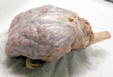
How Does A Sheep Brain Compare To A Human Brain? This in-depth analysis on COMPARE.EDU.VN explores the fascinating similarities and key distinctions between these two mammalian brains. Discover how comparing brain structures sheds light on evolution, function, and potential research avenues using comparative neuroanatomy, brain size, and cognitive capabilities.
1. Introduction: Unveiling the Mysteries of the Brain
The brain, the control center of the body, has always been a subject of immense curiosity. Understanding its structure and function is crucial for comprehending behavior, cognition, and neurological disorders. While studying the human brain directly poses significant challenges, comparative neuroanatomy offers valuable insights. By examining the brains of other animals, particularly mammals like sheep, we can gain a deeper understanding of the human brain’s complexity and evolution. This article on COMPARE.EDU.VN delves into a detailed comparison of the sheep brain and the human brain, highlighting their similarities, differences, and the implications for neuroscience research.
2. Why Compare a Sheep Brain to a Human Brain?
Comparing a sheep brain to a human brain might seem unusual at first, but there are several compelling reasons for this comparison:
- Similarities in Structure: Sheep brains share many structural similarities with human brains, making them a useful model for studying human brain anatomy.
- Availability and Ethics: Sheep brains are readily available from slaughterhouses, making them an accessible and ethically sound alternative to human brain specimens.
- Size and Complexity: While smaller than human brains, sheep brains are complex enough to demonstrate key brain structures and functions.
- Research Applications: Sheep brains have been used in various research areas, including studies of neurodegenerative diseases and brain injury.
3. Brain Size and Overall Structure: A Comparative Overview
3.1. Brain Size
One of the most obvious differences between the sheep brain and the human brain is size. The average human brain weighs about 1300-1400 grams and has a volume of around 1260 cubic centimeters. In contrast, the average sheep brain weighs approximately 140 grams and has a volume of about 140 cubic centimeters. This significant difference in size reflects the greater complexity and cognitive abilities of the human brain.
3.2. Overall Structure
Despite the size difference, both the sheep brain and the human brain share the same basic structural components:
- Cerebrum: The largest part of the brain, responsible for higher-level cognitive functions such as reasoning, memory, and language.
- Cerebellum: Located at the back of the brain, primarily responsible for motor control, coordination, and balance.
- Brainstem: Connects the brain to the spinal cord, regulating essential functions such as breathing, heart rate, and sleep-wake cycles.
Within these major regions, both brains also contain similar structures such as the cerebral cortex, thalamus, hypothalamus, hippocampus, amygdala, and various lobes (frontal, parietal, temporal, and occipital).
4. Key Brain Regions: A Detailed Comparison
4.1. Cerebrum: The Seat of Higher Cognition
The cerebrum is the largest part of the brain and is responsible for many of our higher-level cognitive functions. In both sheep and humans, the cerebrum is divided into two hemispheres, connected by a bundle of nerve fibers called the corpus callosum.
4.1.1. Cerebral Cortex
The cerebral cortex is the outer layer of the cerebrum and is responsible for processing sensory information, planning and executing movements, and higher-level cognitive functions such as language and reasoning. The human cerebral cortex is significantly larger and more convoluted than the sheep cerebral cortex, allowing for greater surface area and more complex neural connections.
- Gyri and Sulci: The convolutions of the cerebral cortex, known as gyri (ridges) and sulci (grooves), increase the surface area of the cortex without increasing the overall volume of the brain. Humans have a much higher density of gyri and sulci compared to sheep, indicating a greater capacity for information processing.
4.1.2. Lobes of the Cerebrum
The cerebrum is divided into four lobes: frontal, parietal, temporal, and occipital. Each lobe is responsible for specific functions, and the relative size and complexity of these lobes differ between sheep and humans.
- Frontal Lobe: The frontal lobe is the largest lobe in the human brain and is responsible for higher-level cognitive functions such as planning, decision-making, working memory, and personality. The frontal lobe is proportionately smaller in sheep, reflecting their less complex cognitive abilities.
- Parietal Lobe: The parietal lobe is responsible for processing sensory information such as touch, temperature, pain, and spatial awareness. The parietal lobe is relatively similar in size and function between sheep and humans.
- Temporal Lobe: The temporal lobe is responsible for processing auditory information, memory, and language. The temporal lobe is also relatively similar in size and function between sheep and humans.
- Occipital Lobe: The occipital lobe is responsible for processing visual information. The occipital lobe is relatively similar in size and function between sheep and humans.
4.2. Cerebellum: The Master of Motor Coordination
The cerebellum is located at the back of the brain and is primarily responsible for motor control, coordination, and balance. While the cerebellum is smaller than the cerebrum in both sheep and humans, it plays a crucial role in coordinating movements and maintaining posture.
4.2.1. Arbor Vitae
The arbor vitae, meaning “tree of life” in Latin, is a distinctive feature of the cerebellum. It is a branching pattern of white matter that resembles a tree. The arbor vitae is present in both sheep and human cerebellums, and it plays a crucial role in transmitting information between the cerebellum and other parts of the brain.
4.2.2. Cerebellar Cortex
The cerebellar cortex is the outer layer of the cerebellum and is responsible for processing sensory information and coordinating movements. The cerebellar cortex is highly folded, forming numerous ridges called folia, which increase the surface area of the cortex.
4.3. Brainstem: The Life Support System
The brainstem connects the brain to the spinal cord and is responsible for regulating essential functions such as breathing, heart rate, blood pressure, and sleep-wake cycles. The brainstem is a relatively similar size and structure in both sheep and humans, reflecting the importance of these basic life-sustaining functions.
4.3.1. Key Structures of the Brainstem
The brainstem consists of several key structures, including the midbrain, pons, and medulla oblongata.
- Midbrain: The midbrain is involved in motor control, vision, hearing, and arousal.
- Pons: The pons is involved in motor control, sensory information, and sleep-wake cycles.
- Medulla Oblongata: The medulla oblongata is responsible for regulating vital functions such as breathing, heart rate, and blood pressure.
4.4. Other Key Brain Structures
In addition to the cerebrum, cerebellum, and brainstem, several other key brain structures are present in both sheep and humans.
4.4.1. Thalamus
The thalamus is a relay station for sensory information, transmitting information from the sensory organs to the cerebral cortex.
4.4.2. Hypothalamus
The hypothalamus regulates essential functions such as body temperature, hunger, thirst, and sleep-wake cycles.
4.4.3. Hippocampus
The hippocampus is involved in memory formation and spatial navigation.
4.4.4. Amygdala
The amygdala is involved in processing emotions such as fear, anger, and pleasure.
5. Functional Differences: How Brain Structure Impacts Behavior
While the sheep brain and the human brain share many structural similarities, there are also significant functional differences that reflect the different cognitive abilities of these two species.
5.1. Cognitive Abilities
Humans possess a much wider range of cognitive abilities compared to sheep, including:
- Language: Humans have the unique ability to communicate using complex language.
- Reasoning: Humans are capable of abstract thought, problem-solving, and logical reasoning.
- Planning: Humans can plan for the future and make decisions based on long-term goals.
- Self-awareness: Humans have a sense of self and are aware of their own thoughts and feelings.
These advanced cognitive abilities are largely due to the greater size and complexity of the human cerebral cortex, particularly the frontal lobe.
5.2. Sensory Perception
While both sheep and humans have similar sensory systems, there are some differences in how they perceive the world.
- Vision: Sheep have excellent peripheral vision, allowing them to detect predators from a wide angle. However, their depth perception is not as good as humans.
- Hearing: Sheep have excellent hearing, allowing them to detect sounds from a long distance.
- Smell: Sheep have a highly developed sense of smell, which they use to find food and identify other sheep.
5.3. Motor Skills
Humans have a much greater range of motor skills compared to sheep, including fine motor skills such as writing and playing musical instruments. This is due to the greater complexity of the human motor cortex and the cerebellum.
6. Evolutionary Considerations: Tracing the Development of the Brain
Comparing the sheep brain and the human brain provides valuable insights into the evolution of the brain. Both sheep and humans are mammals, and their brains share a common evolutionary history. However, over millions of years, the human brain has evolved to become significantly larger and more complex than the sheep brain.
6.1. Brain Size and Encephalization
Encephalization refers to the increase in brain size relative to body size. Humans have a much higher encephalization quotient (EQ) compared to sheep, indicating that their brains are significantly larger than expected for their body size. This increase in brain size has allowed for the development of more complex cognitive abilities.
6.2. Cortical Folding
The degree of cortical folding, as measured by the density of gyri and sulci, is also an indicator of brain complexity. Humans have a much higher density of gyri and sulci compared to sheep, allowing for greater surface area and more complex neural connections.
6.3. Brain Regions and Specialization
The relative size and specialization of different brain regions have also changed over the course of evolution. In humans, the frontal lobe has expanded significantly, allowing for the development of advanced cognitive functions such as planning, decision-making, and language.
7. Research Applications: Utilizing Sheep Brains in Neuroscience
Sheep brains have been used in various research areas, providing valuable insights into brain anatomy, function, and disease.
7.1. Anatomical Studies
Sheep brains are often used in introductory neuroscience courses to teach students about brain anatomy. The sheep brain is large enough to allow students to easily identify key brain structures, and it is also relatively inexpensive and readily available.
7.2. Physiological Studies
Sheep brains have been used to study various physiological processes, such as the effects of drugs on brain activity and the mechanisms of learning and memory.
7.3. Disease Modeling
Sheep brains have been used as models for studying neurodegenerative diseases such as Alzheimer’s disease and Parkinson’s disease. Sheep can develop age-related cognitive decline, making them a useful model for studying the aging brain.
8. Dissection Guide: Exploring the Sheep Brain Firsthand
For those interested in exploring the sheep brain firsthand, a dissection can provide a valuable learning experience. Here’s a step-by-step guide to dissecting a sheep brain:
- Gather your materials: You will need a preserved sheep brain, a dissection tray, a scalpel, scissors, probes, and gloves.
- Observe the external features: Before dissecting the brain, take some time to observe its external features, such as the cerebrum, cerebellum, and brainstem.
- Remove the dura mater: The brain is covered by a tough outer membrane called the dura mater. Carefully remove the dura mater to expose the brain’s surface.
- Identify the lobes of the cerebrum: Identify the frontal, parietal, temporal, and occipital lobes of the cerebrum.
- Locate the major sulci and gyri: Locate the major sulci (grooves) and gyri (ridges) on the surface of the cerebrum.
- Cut the brain in half: Using a scalpel, carefully cut the brain in half along the longitudinal fissure, which separates the two hemispheres.
- Observe the internal structures: Observe the internal structures of the brain, such as the corpus callosum, thalamus, hypothalamus, hippocampus, and amygdala.
- Examine the cerebellum: Examine the cerebellum, noting the arbor vitae (tree of life) and the cerebellar cortex.
- Identify the brainstem structures: Identify the midbrain, pons, and medulla oblongata in the brainstem.
- Dispose of the brain properly: Dispose of the brain according to your local regulations.
9. Ethical Considerations: Respecting Animal Life in Research and Education
It is important to consider the ethical implications of using animal brains in research and education. While sheep brains are readily available from slaughterhouses, it is important to treat them with respect and to minimize any potential suffering.
9.1. The 3Rs Principle
The 3Rs principle, which stands for Replacement, Reduction, and Refinement, provides a framework for ethical animal research.
- Replacement: Replace the use of animals with non-animal methods whenever possible.
- Reduction: Reduce the number of animals used in research to the minimum necessary.
- Refinement: Refine experimental procedures to minimize any potential pain or distress to the animals.
9.2. Alternatives to Dissection
There are several alternatives to dissecting animal brains, such as virtual dissections and computer simulations. These alternatives can provide a valuable learning experience without the need to use animal specimens.
10. The Future of Comparative Neuroanatomy: New Technologies and Discoveries
Comparative neuroanatomy is a rapidly evolving field, with new technologies and discoveries constantly expanding our understanding of the brain.
10.1. Advanced Imaging Techniques
Advanced imaging techniques such as magnetic resonance imaging (MRI) and diffusion tensor imaging (DTI) allow us to visualize the brain’s structure and function in unprecedented detail. These techniques can be used to compare the brains of different species and to identify subtle differences in brain structure and connectivity.
10.2. Genetic Analysis
Genetic analysis can provide insights into the evolutionary relationships between different species and the genetic basis of brain development. By comparing the genomes of different species, we can identify genes that are involved in brain size, structure, and function.
10.3. Computational Modeling
Computational modeling allows us to simulate the brain’s activity and to test hypotheses about brain function. By creating computer models of the brain, we can gain a better understanding of how the brain processes information and how different brain regions interact with each other.
11. Conclusion: A Journey into the Brain’s Complexity
Comparing the sheep brain to the human brain provides a fascinating glimpse into the complexity of the brain and the evolutionary history of cognition. While the human brain is significantly larger and more complex than the sheep brain, both brains share the same basic structural components and perform many of the same functions. By studying the similarities and differences between these two brains, we can gain a deeper understanding of the human brain and the biological basis of behavior.
Whether you’re a student, a researcher, or simply curious about the brain, exploring the world of comparative neuroanatomy can be a rewarding and enlightening experience. The journey to understand the brain is ongoing, and new discoveries are constantly being made. By embracing new technologies and approaches, we can continue to unravel the mysteries of the brain and unlock its full potential.
Unlock deeper comparisons and make informed decisions by visiting COMPARE.EDU.VN today. Our comprehensive resources offer detailed analyses and objective evaluations to guide you every step of the way.
Address: 333 Comparison Plaza, Choice City, CA 90210, United States
Whatsapp: +1 (626) 555-9090
Website: compare.edu.vn
12. Frequently Asked Questions (FAQ)
1. What are the main differences between a sheep brain and a human brain?
The main differences lie in size and complexity. Human brains are much larger and have a more convoluted cerebral cortex, allowing for greater cognitive abilities.
2. Why are sheep brains used in dissection?
Sheep brains are readily available, ethically sourced, and share structural similarities with human brains, making them ideal for educational dissections.
3. What is the dura mater?
The dura mater is a tough outer covering that protects the brain.
4. What is the corpus callosum?
The corpus callosum is a bundle of nerve fibers that connects the two hemispheres of the cerebrum.
5. What is the cerebellum responsible for?
The cerebellum is primarily responsible for motor control, coordination, and balance.
6. What is the brainstem responsible for?
The brainstem regulates essential functions such as breathing, heart rate, and blood pressure.
7. What are the lobes of the cerebrum?
The cerebrum is divided into four lobes: frontal, parietal, temporal, and occipital.
8. What is the function of the frontal lobe?
The frontal lobe is responsible for higher-level cognitive functions such as planning, decision-making, and working memory.
9. What is encephalization?
Encephalization refers to the increase in brain size relative to body size.
10. How can I learn more about brain anatomy?
You can learn more about brain anatomy through textbooks, online resources, and hands-on activities such as brain dissection.
