The remarkable cognitive abilities of dolphins, including self-recognition, complex problem-solving, and intricate communication, have long fascinated scientists. These feats raise a fundamental question: how do dolphin brains compare to human brains, particularly in regions associated with higher-order thinking? This article delves into a comparative analysis of human and dolphin brains, focusing on the prefrontal cortex (PFC), a brain area crucial for advanced cognitive functions.
Simplified comparison of brain evolution in dolphins and humans, highlighting the PFC (orange).
Unraveling the Dolphin Brain’s Mystery: The Prefrontal Cortex
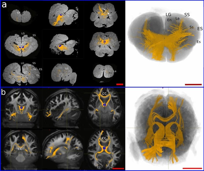
The PFC in humans is well-defined and extensively studied. However, identifying a comparable region in dolphins has proven challenging due to ethical limitations on invasive research. Non-invasive techniques, such as diffusion-weighted imaging (DWI), offer a viable solution. Constrained spherical deconvolution (CSD), a specific DWI technique, allows researchers to map complex fiber pathways in the brain, even in post-mortem specimens.
Mapping the Connections: A Comparative Approach
By applying CSD to both dolphin and human brains, researchers can compare the connectivity patterns originating from the medio-dorsal thalamic nucleus (MDN), a key relay station connected to the PFC. In humans, the MDN projects strongly to the PFC, consistent with its role in higher cognitive functions. In dolphins, similar projections were found leading to a region in the cranio-lateral, ectolateral, and opercular gyri, suggesting a potential PFC homologue.
Similarities and Differences: Evolutionary Considerations
While the connectivity patterns between the MDN and the putative PFC in dolphins show striking similarities to those in humans, there are also notable differences in brain topography. The dolphin’s PFC appears more laterally positioned compared to the human brain, likely due to evolutionary adaptations for aquatic life. The telescoping process in dolphins, where the skull bones rearranged to accommodate the blowhole, may have influenced the brain’s overall shape and the positioning of different cortical areas.
Beyond Structure: Exploring Function and Neurochemistry
While CSD provides crucial insights into structural connectivity, further research is necessary to understand the functional and neurochemical characteristics of the putative dolphin PFC. Investigating the presence of dopaminergic neurons, known to play a significant role in the mammalian PFC, could offer further clues. Advanced imaging techniques, such as functional MRI (fMRI) and functional near-infrared spectroscopy (fNIRS), may also contribute to unraveling the functional mysteries of the dolphin brain.
Conclusion
Comparing human and dolphin brains using advanced imaging techniques like CSD reveals compelling similarities in the connectivity patterns associated with higher cognitive functions. While topographical differences exist due to evolutionary pressures, the presence of a putative PFC in dolphins highlights the complex cognitive capabilities of these marine mammals. Continued research into the functional and neurochemical aspects of the dolphin brain promises to deepen our understanding of intelligence and its evolution across different species.