Do humans have smaller erector spinae compared to apes? This question delves into the fascinating evolutionary adaptations that have shaped our species, and COMPARE.EDU.VN provides a detailed analysis of these anatomical differences. By exploring the spinal musculature of humans and apes, we can gain a deeper understanding of the musculoskeletal variations that enabled bipedalism. We’ll explore the comparative anatomy, evolutionary perspectives, and functional implications. Key terms include spinal adaptations, bipedal locomotion, and human evolution.
1. Introduction: Exploring the Evolutionary Divide
The transition to bipedalism marks a significant milestone in human evolution, distinguishing our ancestors from other primates. Understanding how our anatomy has evolved to support upright walking and balance is crucial. This exploration begins with a look at the erector spinae muscles, essential components of our spinal structure. These muscles play a pivotal role in maintaining posture and facilitating movement.
The objective is to clarify whether humans have smaller erector spinae muscles compared to apes and, if so, why this difference exists. We will examine the anatomy of these muscles in both humans and apes, considering their functions and how they contribute to overall spinal health and stability. This comparison is essential to understanding the unique evolutionary trajectory of humans.
2. Defining the Erector Spinae: Anatomy and Function
2.1. What are the Erector Spinae Muscles?
The erector spinae muscles are a group of muscles located along the vertebral column, extending from the sacrum to the skull. They consist of three primary muscles: iliocostalis, longissimus, and spinalis.
- Iliocostalis: This muscle is the most lateral of the group, running from the iliac crest to the ribs and transverse processes of the vertebrae.
- Longissimus: Situated between the iliocostalis and spinalis, the longissimus extends from the sacrum to the skull, attaching to the transverse processes of the vertebrae and ribs.
- Spinalis: The most medial of the erector spinae muscles, the spinalis runs along the spinous processes of the vertebrae.
2.2. Key Functions of the Erector Spinae
The erector spinae muscles perform several critical functions:
- Extension of the Spine: These muscles are primarily responsible for extending the vertebral column, allowing us to stand upright and arch our backs.
- Lateral Flexion: They assist in lateral flexion, enabling us to bend to the side.
- Rotation: The erector spinae muscles also contribute to spinal rotation, although their role is less pronounced than that of other muscles like the rotatores and multifidus.
- Posture Maintenance: They play a vital role in maintaining posture by counteracting the forces of gravity and supporting the trunk.
- Control of Spinal Movements: These muscles provide stability and control during various movements, such as walking, running, and lifting.
Understanding the anatomy and functions of the erector spinae muscles is crucial for comparing their characteristics in humans and apes. This knowledge forms the foundation for exploring the evolutionary adaptations that have shaped our spinal structures.
3. Comparative Anatomy: Humans vs. Apes
3.1. Erector Spinae Size and Insertion Points
Comparing the size and insertion points of the erector spinae muscles in humans and apes provides insights into the functional differences between these species. While apes generally have larger and more robust erector spinae muscles, humans exhibit unique adaptations tailored to bipedalism.
- Size Comparison:
- Apes: Apes, particularly great apes like gorillas and chimpanzees, have proportionally larger erector spinae muscles compared to humans. This greater size is associated with their need for powerful trunk stabilization during quadrupedal locomotion and climbing.
- Humans: Humans have relatively smaller erector spinae muscles. This reduction in size may be an adaptation to reduce energy expenditure during bipedalism.
- Insertion Points:
- Apes: In apes, the erector spinae muscles have broad insertion points along the iliac crest and sacrum, providing a wide base for muscle attachment. This extensive attachment area supports powerful trunk movements and stability.
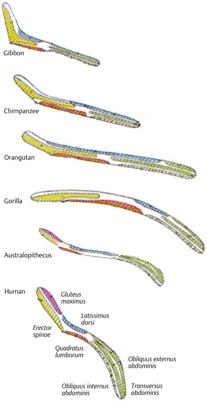
- Humans: Humans also have insertion points along the iliac crest and sacrum, but the area is generally smaller compared to apes. The gluteus maximus muscle originates high on the pelvis at the level of the iliac crest, enhancing the capacity for trunk erection. The muscles balancing the trunk originate from the iliac crest. These include the oblique and transverse abdominal muscles, latissimus dorsi, quadratus lumborum, trunk erectors, and gluteus maximus.
3.2. Spinal Curvature and Posture
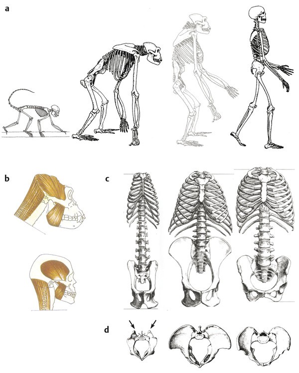
Differences in spinal curvature and posture further distinguish humans from apes. Humans possess a unique S-shaped spine with distinct cervical, thoracic, lumbar, and sacral curves, while apes have a more C-shaped spine.
- Spinal Curvature:
- Apes: Apes have a kyphotic thoracolumbar curvature, which means their spine is curved outward in the thoracic and lumbar regions. This curvature is suited for quadrupedal locomotion.
- Humans: Humans have a double S-shaped spine, including a cervical lordosis (inward curve in the neck), a thoracic kyphosis (outward curve in the upper back), a lumbar lordosis (inward curve in the lower back), and a sacral kyphosis (outward curve in the sacrum). The lumbar lordosis brings the center of body weight closer to the hip joint, minimizing muscular work to maintain equilibrium.
- Posture:
- Apes: Apes often exhibit a flexed hip and knee posture, which shifts their center of gravity forward. This posture requires strong erector spinae muscles to counteract the forward lean and maintain balance.
- Humans: Humans maintain an upright posture with extended hips and knees, placing the center of gravity directly over the feet. This posture reduces the demand on the erector spinae muscles for balance.
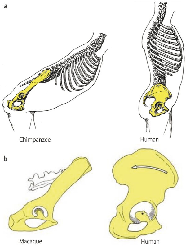
3.3. Pelvic Structure
The structure of the pelvis also differs significantly between humans and apes, impacting the function of the erector spinae muscles.
- Pelvic Shape:
- Apes: Apes have a long and narrow pelvis with laterally flaring iliac blades. The distance between the hip joint and sacroiliac joint is relatively long, increasing rotational moments of the iliac segment.
- Humans: Humans have a shorter and broader pelvis with frontally oriented iliac blades. The approximation between the sacroiliac and hip joints reduces the rotational moments, diminishing the muscular work needed to maintain equilibrium. The gluteus maximus muscle originates high on the pelvis, enhancing its capacity for trunk erection.
- Muscle Attachments:
- Apes: Apes have an extensive area of origin for the latissimus dorsi and quadratus lumborum, important muscles for supporting the trunk and pelvis during arm suspension.
- Humans: Humans have an anteriorly curved lateral section of the iliac blades, modifying the position of the lesser gluteal muscles. This configuration implies a different mechanism of lateral pelvic balance during bipedalism.
3.4. Table Summarizing Key Anatomical Differences
| Feature | Apes | Humans |
|---|---|---|
| Erector Spinae Size | Larger, more robust | Relatively smaller |
| Spinal Curvature | Kyphotic thoracolumbar curvature (C-shaped) | Double S-shaped (cervical lordosis, thoracic kyphosis, lumbar lordosis, sacral kyphosis) |
| Pelvic Shape | Long and narrow, laterally flaring iliac blades | Shorter and broader, frontally oriented iliac blades |
| Posture | Flexed hip and knee posture | Upright posture with extended hips and knees |
| Muscle Attachments | Extensive area of origin for latissimus dorsi and quadratus lumborum | Anteriorly curved lateral section of the iliac blades, modifying the position of the lesser gluteal muscles |
| Sacroiliac Distance | Long distance between the hip joint and sacroiliac joint | Shorter distance between the hip joint and sacroiliac joint |
These anatomical differences highlight the adaptations that have shaped human bipedalism, contrasting with the quadrupedal and climbing adaptations of apes.
4. Evolutionary Perspectives on Erector Spinae Development
4.1. Transition to Bipedalism
The transition from quadrupedalism to bipedalism is a defining event in human evolution. This shift required significant anatomical changes, particularly in the spine and pelvis. The development of the erector spinae muscles played a crucial role in supporting upright posture and movement.
- Early Hominids: Early hominids, such as Australopithecus afarensis, exhibited a mosaic of ape-like and human-like features. Their erector spinae muscles were likely larger than those of modern humans, reflecting the need for greater trunk stabilization during both arboreal and terrestrial locomotion.
- Evolutionary Pressures: As hominids became more terrestrial and adopted bipedalism as their primary mode of locomotion, selective pressures favored adaptations that enhanced efficiency and reduced energy expenditure. Smaller erector spinae muscles may have been advantageous in this context, as they required less energy to maintain posture.
4.2. Spinal Adaptations Over Time
The human spine has undergone significant changes over millions of years, adapting to the demands of bipedalism. These adaptations include the development of spinal curvatures, changes in vertebral segmentation, and modifications in the orientation of facet joints.
- Spinal Curvatures: The evolution of the double S-shaped spine in humans is a key adaptation to bipedalism. The lumbar lordosis, in particular, helps to bring the center of gravity closer to the hips, reducing the amount of muscular effort needed to maintain balance.
- Vertebral Segmentation: The number of lumbar vertebrae also plays a role in spinal flexibility and lordosis. Humans typically have five lumbar vertebrae, which is longer and more flexible than the lumbar spine of great apes.
- Facet Joint Orientation: The orientation of the facet joints at the thoracolumbar junction differs between early hominids and modern humans. Early hominids had a more cranially located transitional vertebra, which may have provided greater rotational stability of the trunk.
4.3. Muscle Development and Energetic Efficiency
The reduction in size of the erector spinae muscles in humans may be linked to the need for energetic efficiency during bipedal locomotion. Smaller muscles require less energy to maintain posture and perform movements, which could have been advantageous in environments where food resources were limited.
- Metabolic Costs: Maintaining large muscle mass is metabolically expensive, requiring significant energy input. By reducing the size of the erector spinae muscles, humans may have reduced their overall metabolic costs, freeing up energy for other activities.
- Locomotor Efficiency: Bipedalism is a more energetically efficient mode of locomotion than quadrupedalism, particularly over long distances. The reduction in size of the erector spinae muscles may have contributed to this efficiency by reducing the amount of energy needed to maintain posture and balance.
4.4. Scenarios for Spinal Segmentation Evolution
Different scenarios have been proposed for the evolution of human spinal segmentation. The short-backed scenario suggests that the short lumbar column of chimpanzees and gorillas is primitive. The long-backed scenario proposes that a six-segment-long lumbar spine was retained in the ancestors of apes and humans.
- Intermediate Scenario: The intermediate scenario argues for five lumbar vertebrae as the primitive condition in great apes and humans. This scenario is supported by evidence from Oreopithecus, a great ape that lived 8 million years ago and also had five lumbar vertebrae. The common ancestor of chimpanzees and humans already had five lumbar vertebrae, which facilitated the adoption of lumbar lordosis and thus of bipedal locomotion.
5. Functional Implications of Erector Spinae Size Differences
5.1. Postural Control and Stability
The size and strength of the erector spinae muscles have significant implications for postural control and stability. While apes rely on larger erector spinae muscles for powerful trunk stabilization, humans have adapted to maintain balance with relatively smaller muscles.
- Apes: Apes require strong erector spinae muscles to counteract the forward lean of their posture and maintain balance during quadrupedal locomotion and climbing. Their larger muscles provide the necessary power and stability for these activities.
- Humans: Humans rely on a combination of spinal curvatures, pelvic structure, and muscle activation patterns to maintain upright posture and balance. While their erector spinae muscles are smaller, they are still essential for providing stability and controlling spinal movements.
5.2. Locomotion and Movement Patterns
The differences in erector spinae size also affect locomotion and movement patterns. Apes use their strong erector spinae muscles to propel themselves forward during quadrupedal walking and to stabilize their trunks during climbing. Humans use their smaller erector spinae muscles to maintain balance and control spinal movements during bipedal walking, running, and other activities.
- Apes: Apes exhibit a more flexed hip and knee posture during locomotion, which requires greater activation of the erector spinae muscles to maintain balance. Their powerful trunk muscles allow them to navigate complex arboreal environments.
- Humans: Humans maintain an upright posture with extended hips and knees during locomotion, which reduces the demand on the erector spinae muscles. Their spinal curvatures and pelvic structure help to distribute weight and minimize muscular effort.
5.3. Risk of Spinal Injuries
The size and strength of the erector spinae muscles may also influence the risk of spinal injuries. Weak erector spinae muscles can contribute to poor posture, muscle imbalances, and increased susceptibility to back pain and injuries.
- Apes: While apes have strong erector spinae muscles, they are still at risk of spinal injuries, particularly if they engage in strenuous activities or experience trauma. Their larger muscles may provide some protection against injury.
- Humans: Humans are particularly vulnerable to back pain and injuries, due in part to their relatively smaller erector spinae muscles and the stresses of bipedalism. Maintaining good posture, strengthening the erector spinae muscles, and practicing proper lifting techniques can help to reduce the risk of spinal injuries.
5.4. Core Stability and Compensatory Mechanisms
Core stability is essential for maintaining spinal health and preventing injuries. The erector spinae muscles are part of the core musculature, which also includes the abdominal muscles, obliques, and transverse abdominis.
- Core Stability: A strong core helps to stabilize the spine, reduce stress on the erector spinae muscles, and improve overall posture and movement patterns. Engaging in core strengthening exercises can help to improve spinal health and reduce the risk of injuries.
- Compensatory Mechanisms: When the erector spinae muscles are weak or injured, other muscles may compensate to maintain posture and stability. This can lead to muscle imbalances, altered movement patterns, and increased risk of further injury.
6. Implications for Modern Humans: Health and Fitness
6.1. Back Pain and Posture
The relatively smaller size of the erector spinae muscles in modern humans has implications for back pain and posture. Weak erector spinae muscles can contribute to poor posture, muscle imbalances, and increased susceptibility to back pain.
- Poor Posture: Slouching, hunching, and other forms of poor posture can place excessive stress on the erector spinae muscles, leading to fatigue, pain, and injury.
- Muscle Imbalances: Weak erector spinae muscles can lead to muscle imbalances, where other muscles compensate to maintain posture and stability. This can result in altered movement patterns and increased risk of further injury.
- Back Pain: Back pain is a common problem among modern humans, and weak erector spinae muscles are often a contributing factor. Strengthening the erector spinae muscles and improving posture can help to reduce back pain and improve overall spinal health.
6.2. Exercise and Strengthening
Regular exercise and strengthening of the erector spinae muscles can help to improve posture, reduce back pain, and enhance overall spinal health. There are many exercises that can target the erector spinae muscles, including:
- Back Extensions: Back extensions are a simple and effective exercise for strengthening the erector spinae muscles. They can be performed on a back extension machine or using a stability ball.
- Supermans: Supermans involve lying face down and simultaneously lifting the arms and legs off the ground. This exercise targets the erector spinae muscles, glutes, and hamstrings.
- Bird Dogs: Bird dogs involve starting on your hands and knees and simultaneously extending one arm forward and the opposite leg backward. This exercise targets the erector spinae muscles, glutes, and core.
- Deadlifts: Deadlifts are a more advanced exercise that can be highly effective for strengthening the erector spinae muscles. However, they should be performed with proper form to avoid injury.
- Glute Bridges: Glute bridges engage the glutes and erector spinae, improving hip extension and spinal stability.
- Plank: Planks build core strength, indirectly supporting the erector spinae by stabilizing the spine.
6.3. Lifestyle Considerations
Adopting a healthy lifestyle can also help to support spinal health and reduce the risk of back pain. This includes:
- Maintaining a Healthy Weight: Being overweight or obese can place excessive stress on the spine, increasing the risk of back pain and injury.
- Practicing Good Posture: Consciously maintaining good posture throughout the day can help to reduce stress on the erector spinae muscles and improve spinal health.
- Using Proper Lifting Techniques: When lifting heavy objects, it is important to use proper lifting techniques to avoid injury. This includes bending at the knees, keeping the back straight, and holding the object close to the body.
- Staying Active: Regular physical activity can help to strengthen the erector spinae muscles, improve posture, and reduce the risk of back pain.
- Proper Ergonomics: Setting up your workspace to support good posture can reduce strain on your back.
6.4. Professional Guidance
Seeking guidance from a healthcare professional, such as a physical therapist or chiropractor, can be beneficial for addressing back pain and improving spinal health. These professionals can provide individualized recommendations for exercise, posture correction, and other lifestyle modifications.
- Physical Therapy: Physical therapists can help to assess spinal health, identify muscle imbalances, and develop a tailored exercise program to strengthen the erector spinae muscles and improve posture.
- Chiropractic Care: Chiropractors can help to align the spine, reduce nerve irritation, and improve overall spinal function.
7. Conclusion: The Evolutionary Trade-Off
In conclusion, humans do have smaller erector spinae muscles compared to apes. This reduction in size is an evolutionary trade-off that has allowed humans to adapt to bipedalism and improve energetic efficiency. While smaller erector spinae muscles may increase the risk of back pain and injury, regular exercise, good posture, and a healthy lifestyle can help to mitigate these risks.
The evolutionary journey from quadrupedal apes to bipedal humans has resulted in significant changes to our spinal structure and musculature. Understanding these changes is essential for appreciating the unique adaptations that have shaped our species and for addressing the health challenges that arise from these adaptations.
By exploring the comparative anatomy, evolutionary perspectives, and functional implications of erector spinae size differences, we gain a deeper understanding of the musculoskeletal variations that enabled bipedalism. The study of human evolution continues to provide valuable insights into the complexities of our anatomy and the challenges of maintaining spinal health in the modern world.
For more in-depth comparisons and comprehensive information on evolutionary adaptations, visit COMPARE.EDU.VN, your trusted source for detailed analyses and informed decision-making.
8. Call to Action
Are you looking for detailed comparisons of human and ape anatomy? Do you want to understand the evolutionary adaptations that have shaped our bodies? Visit COMPARE.EDU.VN today to explore our comprehensive articles and make informed decisions about your health and fitness.
Need more information or have specific questions? Contact us at:
- Address: 333 Comparison Plaza, Choice City, CA 90210, United States
- WhatsApp: +1 (626) 555-9090
- Website: COMPARE.EDU.VN
9. Frequently Asked Questions (FAQ)
Q1: Why are the erector spinae muscles important?
The erector spinae muscles are essential for maintaining posture, extending the spine, and supporting movement.
Q2: Do apes have stronger backs than humans?
Apes generally have larger and more robust erector spinae muscles, but humans have adapted unique strategies for stability.
Q3: How can I strengthen my erector spinae muscles?
Exercises like back extensions, supermans, and deadlifts can help strengthen the erector spinae muscles.
Q4: What role does posture play in back health?
Good posture helps to reduce stress on the erector spinae muscles and improve spinal health.
Q5: Can weak erector spinae muscles cause back pain?
Yes, weak erector spinae muscles can contribute to poor posture and increased susceptibility to back pain.
Q6: What is the lumbar lordosis, and why is it important?
The lumbar lordosis is the inward curve in the lower back, which helps to bring the center of gravity closer to the hips and reduce muscular effort.
Q7: How does the pelvic structure differ between humans and apes?
Humans have a shorter and broader pelvis compared to the long and narrow pelvis of apes.
Q8: Is bipedalism more energetically efficient than quadrupedalism?
Yes, bipedalism is more energetically efficient, particularly over long distances.
Q9: What lifestyle changes can I make to improve my spinal health?
Maintaining a healthy weight, practicing good posture, and staying active can help improve your spinal health.
Q10: Where can I find more detailed comparisons of human and ape anatomy?
Visit compare.edu.vn for comprehensive articles and detailed analyses.
Q11: What is core stability and how does it relate to erector spinae?
Core stability refers to the strength and coordination of the muscles surrounding the trunk and spine, including the erector spinae, abdominals, and obliques. A strong core helps to stabilize the spine, reduce stress on the erector spinae, and improve overall posture and movement patterns.
Q12: What are the risks of having weak erector spinae muscles?
Weak erector spinae muscles can contribute to poor posture, muscle imbalances, increased susceptibility to back pain and injuries, and reduced overall spinal stability.
alt: Skeletal comparison showing differences in body proportions and head posture between a quadrupedal monkey, a knuckle-walking gorilla, and a modern human, highlighting key anatomical adaptations for locomotion and balance.
alt: Diagram illustrating the muscle origins on the iliac crest in great apes and hominids, emphasizing the extensive area of origin for latissimus dorsi and quadratus lumborum in apes, compared to the gluteus maximus origin on the ilium in hominids.
alt: Comparative illustration depicting the spinal curvature and pelvis differences between nonhuman primates with a single thoracolumbar curve and humans with a lumbar lordosis that helps bring the center of body weight closer to the hip joint.