When Comparing Prokaryotes And Eukaryotes Flagella, the differences lie in their structure, mechanism of movement, and energy source; COMPARE.EDU.VN offers detailed comparisons illuminating these distinctions. Understanding these variances is crucial for comprehending the diverse strategies cells use for motility and interaction with their environments, enhancing knowledge in cell biology and microbiology with comparisons of cellular components and locomotive appendages.
1. Comparing Prokaryotic and Eukaryotic Flagella: A Detailed Overview
Prokaryotes, including bacteria and archaea, represent the earliest forms of life, with eukaryotes thought to have evolved approximately 2.7 billion years ago. Eukaryotic evolution is believed to have originated from a symbiotic relationship between two prokaryotes that merged through endosymbiosis. This merger led to the development of membrane-bound organelles like mitochondria, providing eukaryotes with the energy needed to evolve into more complex cells. Recent research, however, has challenged these long-held beliefs, showing that some prokaryotic bacteria can “eat” other cells, contradicting the assumption that only eukaryotes perform endocytosis. This requires a reevaluation of the theories surrounding the origins of eukaryotes.
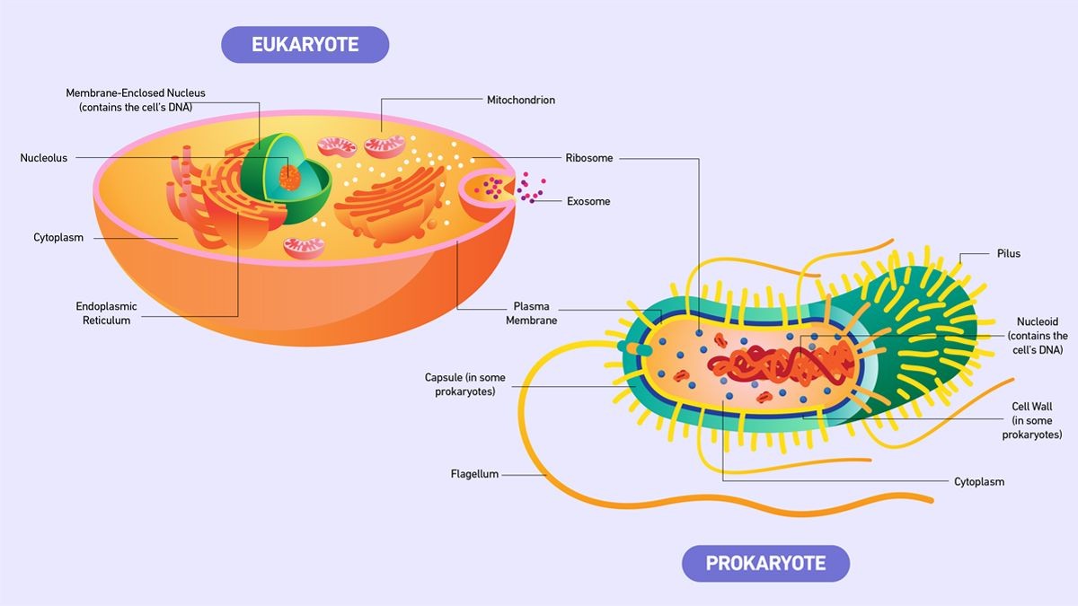
The primary distinction between prokaryotic and eukaryotic cells lies in the presence of a membrane-bound nucleus in eukaryotes, which houses genetic information. In prokaryotes, DNA is bundled together in the nucleoid region without a surrounding membrane. Eukaryotes possess numerous membrane-bound organelles, which are absent in prokaryotes. Eukaryotic DNA is organized into multiple molecules of double-stranded linear DNA within the nucleus, while prokaryotic DNA is typically double-stranded, circular, and located in the cytoplasm, although exceptions with linear plasmids and chromosomes exist.
2. Key Similarities Between Prokaryotes and Eukaryotes
Despite significant differences, prokaryotic and eukaryotic cells share four fundamental features:
- DNA: Both cell types use DNA as their genetic material.
- Plasma Membrane: A cell membrane encloses both cell types, acting as a barrier between the interior and exterior environment.
- Cytoplasm: Both have cytoplasm, the gel-like substance filling the cell.
- Ribosomes: Both cells utilize ribosomes for protein synthesis.
3. What Are the Main Structural and Functional Differences Between Prokaryotes and Eukaryotes?
Prokaryotes and eukaryotes differ substantially in their structure and function. Key differences are summarized in Table 1.
Table 1: Differences Between Prokaryotes and Eukaryotes
| Feature | Prokaryote | Eukaryote |
|---|---|---|
| Nucleus | Absent | Present |
| Membrane-bound Organelles | Absent | Present |
| Cell Structure | Unicellular | Mostly multicellular; some unicellular |
| Cell Size | Typically smaller (0.1–5 μm); however, a much larger (centimeter-long) bacterium was recently discovered in a mangrove swamp. | Larger (10–100 μm) |
| Complexity | Simpler | More complex |
| DNA Form | Often circular; however, linear plasmids and chromosomes have been found in certain prokaryotes. | Linear |
| Examples | Bacteria, archaea | Animals, plants, fungi, protists |
| Flagella Structure | Simple, composed of flagellin; moves by rotation. | Complex, composed of microtubules; moves in a whip-like fashion. |
| Energy Source for Flagella | Proton motive force. | ATP (adenosine triphosphate). |
| Transcription/Translation | Coupled; translation begins during mRNA synthesis. | Not coupled; transcription occurs in the nucleus, translation in the cytoplasm. |

3.1. Transcription and Translation in Prokaryotes vs Eukaryotes
In prokaryotic cells, transcription and translation are closely coupled, meaning translation starts while mRNA is being synthesized. In contrast, eukaryotic cells separate transcription and translation. Transcription occurs in the nucleus, where mRNA is produced, then the mRNA exits the nucleus, and translation occurs in the cytoplasm.
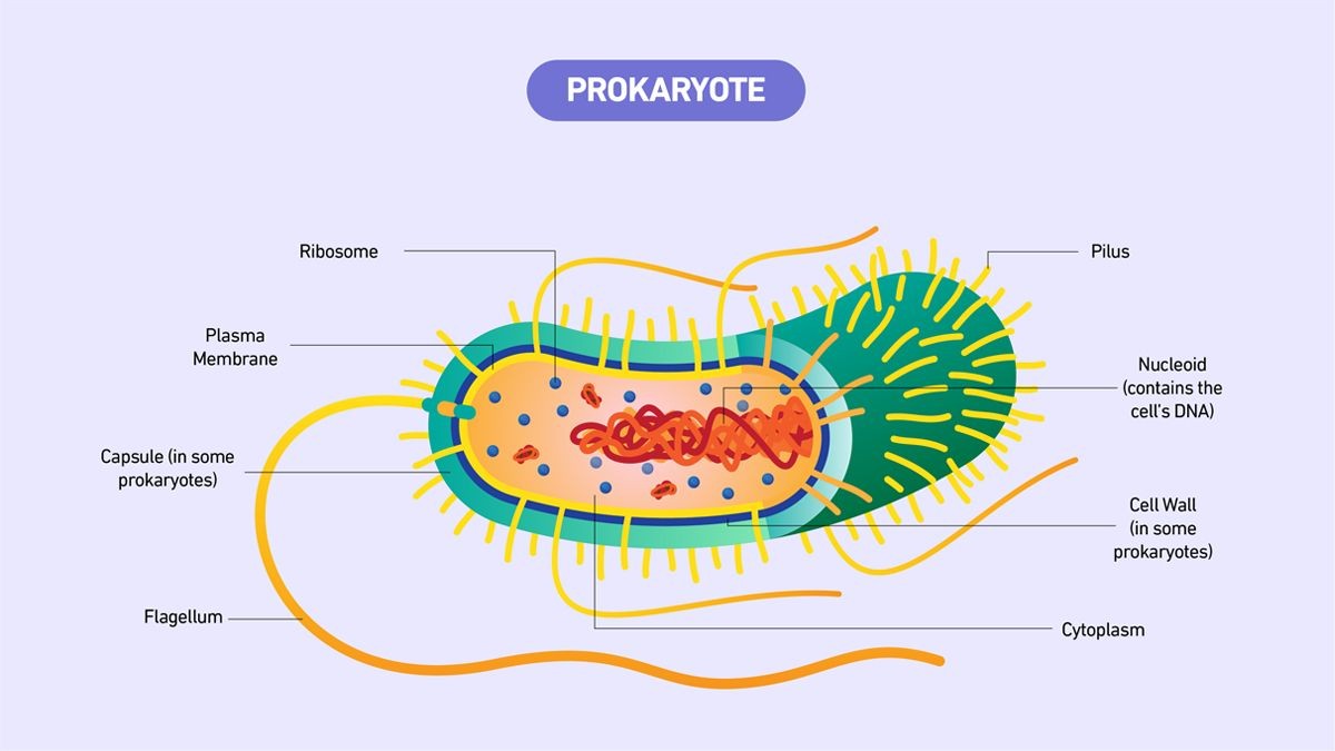
4. What Defines a Prokaryote?
Prokaryotes are divided into two domains: bacteria and archaea. They are unicellular organisms lacking membrane-bound structures. Prokaryotic cells are typically small and simple, ranging from 0.1–5 μm in diameter.
4.1. Prokaryotic Cell Features
A typical prokaryotic bacterial cell includes these key components:
- Nucleoid: A central region containing the cell’s DNA.
- Ribosome: Responsible for protein synthesis.
- Cell Wall: Provides structure and protection. Most bacteria have a rigid cell wall made of peptidoglycans (carbohydrates and proteins).
- Cell Membrane: Separates the cell from the external environment.
- Capsule: A carbohydrate layer surrounding the cell wall in some bacteria, aiding in attachment and protection.
- Pili: Rod-shaped structures involved in attachment and DNA transfer.
- Flagella: Tail-like structures assisting in movement.
4.2. Examples of Prokaryotes
Bacteria and archaea are the two primary types of prokaryotes.
4.3. Do Prokaryotes Have a Nucleus?
Prokaryotes lack a nucleus. Their DNA is located in the nucleoid, a central region without a membrane. Prokaryotic DNA typically consists of a single circular chromosome. They also lack other membrane-bound structures like the endoplasmic reticulum.
4.4. Do Prokaryotes Have Mitochondria?
Prokaryotes do not have mitochondria. Mitochondria are exclusive to eukaryotic cells, along with other membrane-bound structures such as the nucleus and the Golgi apparatus.
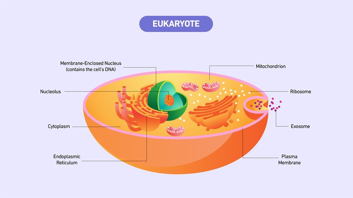
5. What Defines a Eukaryote?
Eukaryotes are organisms with cells containing a nucleus and other organelles enclosed by a plasma membrane. Organelles are internal structures performing various functions, including energy production and protein synthesis.
Eukaryotic cells are large (around 10–100 μm) and complex. While most eukaryotes are multicellular, some are single-celled organisms.
5.1. Eukaryotic Cell Features
Each membrane-bound structure within a eukaryotic cell performs specific cellular functions:
- Nucleus: Stores genetic information in chromatin form.
- Nucleolus: Produces ribosomal RNA inside the nucleus.
- Plasma Membrane: A phospholipid bilayer surrounding the cell and its organelles.
- Cytoskeleton: Protein fibers that shape the cell and position organelles.
- Cell Wall: Found in plant cells, providing structural support and protection.
- Ribosomes: Responsible for protein synthesis.
- Mitochondria: Responsible for energy production.
- Cytoplasmic Space: The region between the nuclear envelope and plasma membrane.
- Cytoplasm: The total inner-cellular volume, excluding the nucleus, including the cytosol and organelles.
- Cytosol: The gel-like substance in the cytoplasm, excluding the contents of organelles.
- Endoplasmic Reticulum: An organelle dedicated to protein maturation and transportation.
- Vesicles and Vacuoles: Membrane-bound sacs involved in transportation and storage.
Other common organelles include the Golgi apparatus, chloroplasts, and lysosomes.
5.2. Examples of Eukaryotes
Animals, plants, fungi, algae, and protozoans are all eukaryotes.
6. Flagella in Prokaryotes vs Eukaryotes: Unveiling the Structural and Functional Differences
When comparing prokaryotic and eukaryotic flagella, the variations extend beyond mere nomenclature, touching upon the very essence of their construction, operational mechanisms, and the energy sources they harness. Flagella, the locomotive appendages of cells, play a pivotal role in the motility and chemotaxis of organisms across various biological domains.
6.1. Structural Variations in Flagella
- Prokaryotic Flagella: These flagella are characterized by their simplicity and are composed of a protein called flagellin. The prokaryotic flagellum is smaller and simpler than its eukaryotic counterpart. It comprises three main parts:
- Filament: The long, helical structure extending from the cell surface, composed of flagellin subunits.
- Hook: A curved structure connecting the filament to the basal body, allowing it to rotate.
- Basal Body: Embedded in the cell membrane and wall, acting as a motor to drive flagellar rotation.
Alt Text: Diagram illustrating the simple structure of a prokaryotic flagellum, including the filament, hook, and basal body.
- Eukaryotic Flagella: Eukaryotic flagella are structurally more complex, being composed of microtubules arranged in a “9+2” array. The structure includes:
- Axoneme: The core structure consisting of nine pairs of microtubules surrounding a central pair.
- Dynein Arms: Protein structures extending from the microtubules, responsible for the sliding movement that causes the flagellum to bend.
- Basal Body: Similar to centrioles, organizing the microtubules and anchoring the flagellum to the cell.
- Plasma Membrane: An outer membrane covering the flagellum, continuous with the cell membrane.
Alt Text: Diagram showcasing the complex “9+2” microtubule arrangement in a eukaryotic flagellum, with axoneme, dynein arms, and basal body.
6.2. Mechanism of Movement
- Prokaryotic Flagella: These operate like a propeller, rotating to propel the cell through its environment. The rotation is powered by a motor at the base of the flagellum.
- Eukaryotic Flagella: These move in a whip-like fashion, bending and flexing to generate movement. The motion is driven by the sliding of microtubules against each other, powered by ATP.
6.3. Energy Source
- Prokaryotic Flagella: Utilize the proton motive force, an electrochemical gradient across the cell membrane, to power the rotation of the flagellum.
- Eukaryotic Flagella: Depend on ATP (adenosine triphosphate), the cell’s primary energy currency, to fuel the movement of dynein arms and the subsequent bending motion.
6.4. Functional Implications
The structural and functional differences in prokaryotic and eukaryotic flagella reflect the diverse strategies employed by cells for motility. Prokaryotic flagella, with their simple design and rotational mechanism, are well-suited for rapid movement in relatively simple environments. Eukaryotic flagella, on the other hand, with their complex structure and whip-like motion, allow for more precise and controlled movement, essential for navigating complex environments and performing specific tasks, such as moving fluids across cell surfaces.
6.5. Evolutionary Context
The disparities in flagellar structure and function also highlight the evolutionary divergence between prokaryotes and eukaryotes. Prokaryotic flagella represent an early evolutionary adaptation for motility, while eukaryotic flagella are a more recent development, reflecting the increased complexity and sophistication of eukaryotic cells.
7. Detailed Comparison of Flagella Components
To fully grasp the differences between prokaryotic and eukaryotic flagella, let’s delve into a comparative analysis of their components, energy mechanisms, and motion dynamics.
| Feature | Prokaryotic Flagella | Eukaryotic Flagella |
|---|---|---|
| Composition | Primarily flagellin protein | Microtubules (9+2 arrangement), dynein, and other proteins |
| Structure | Simple, consisting of a filament, hook, and basal body | Complex, consisting of an axoneme, dynein arms, basal body, and plasma membrane |
| Motor Location | Basal body, located in the cell membrane | Dynein arms, which slide microtubules past each other |
| Energy Source | Proton motive force (electrochemical gradient) | ATP (adenosine triphosphate) |
| Movement | Rotation (like a propeller) | Whip-like motion (bending and flexing) |
| Size | Smaller | Larger |
| Function | Primarily movement and chemotaxis | Movement, sensory functions, and fluid movement across cell surfaces |
| Regulation | Regulated by environmental signals affecting motor activity | Regulated by complex signaling pathways involving calcium and other factors |
| Assembly | Self-assembly of flagellin subunits | Complex assembly process involving multiple proteins and cellular structures |
| Evolutionary Origin | Thought to have evolved independently in bacteria and archaea | Thought to have evolved from eukaryotic cytoskeletal components |
| Glycosylation | Rare or absent | Common; glycosylation of flagellar proteins is important for their function and stability. |
| Membrane Covering | Lacks a membrane covering; directly exposed to the external environment. | Enclosed by the plasma membrane, which is continuous with the cell membrane. |
| Genetic Control | Encoded by genes organized in operons, allowing coordinated expression of flagellar components. | Encoded by genes scattered throughout the genome, requiring more complex regulatory mechanisms. |
| Role in Disease | Important virulence factor in many pathogenic bacteria, aiding in colonization and invasion of host tissues. | Dysfunction can lead to various developmental and physiological disorders, such as primary ciliary dyskinesia. |
| Target for Drugs | Target for antibacterial drugs aimed at inhibiting bacterial motility and virulence. | Potential target for therapies aimed at correcting ciliary and flagellar defects in human diseases. |
| Environmental Adaptation | Highly adaptable to various environmental conditions, allowing bacteria to thrive in diverse habitats. | Adaptation to specific environments, such as the respiratory tract or reproductive system, where ciliary function is critical. |
8. The Role of Flagella in Different Organisms
Understanding the role of flagella in various organisms provides deeper insights into their significance and functional adaptability.
8.1. Bacteria
In bacteria, flagella are crucial for:
- Motility: Allowing bacteria to move towards nutrients or away from harmful substances.
- Chemotaxis: Guiding bacteria towards chemical signals, such as food sources or host cells.
- Virulence: Aiding pathogenic bacteria in colonizing and infecting host tissues.
- Biofilm Formation: Contributing to the formation of biofilms, communities of bacteria attached to surfaces.
8.2. Archaea
In archaea, flagella (archaella) are essential for:
- Motility: Enabling archaea to move in diverse environments, including extreme conditions.
- Chemotaxis: Guiding archaea towards favorable conditions, such as specific chemical gradients.
- Nutrient Acquisition: Facilitating the acquisition of nutrients in nutrient-poor environments.
- Environmental Adaptation: Supporting adaptation to extreme environments, such as high temperatures or acidic conditions.
8.3. Eukaryotes
In eukaryotes, flagella (or cilia) play various roles, including:
- Motility: Propelling single-celled eukaryotes through water or other fluids.
- Fluid Movement: Moving fluids across cell surfaces, such as in the respiratory tract to clear mucus.
- Sensory Functions: Detecting environmental signals, such as light or chemicals.
- Reproduction: Facilitating the movement of sperm cells in animals.
- Development: Playing a role in embryonic development and tissue organization.
9. Clinical Significance of Flagella
Flagella are not only essential for the survival and function of various organisms but also hold clinical significance, particularly in the context of bacterial infections and human health.
9.1. Bacterial Infections
In pathogenic bacteria, flagella are important virulence factors that contribute to the bacteria’s ability to cause disease:
- Adherence: Flagella can aid bacteria in adhering to host cells and tissues, facilitating colonization.
- Invasion: Flagella can promote the invasion of host tissues, allowing bacteria to spread within the body.
- Biofilm Formation: Flagella can contribute to the formation of biofilms, which can protect bacteria from antibiotics and the host immune system.
- Immune Evasion: Flagella can help bacteria evade the host immune system, allowing them to persist and cause chronic infections.
9.2. Human Health
Dysfunction of eukaryotic flagella (cilia) can lead to various human diseases and disorders:
- Primary Ciliary Dyskinesia (PCD): A genetic disorder characterized by defects in ciliary structure and function, leading to chronic respiratory infections, infertility, and situs inversus (reversal of the left-right asymmetry of organs).
- Infertility: Defects in sperm flagella can cause infertility in males, as the sperm are unable to swim and fertilize the egg.
- Respiratory Diseases: Impaired ciliary function in the respiratory tract can lead to chronic respiratory infections, such as bronchitis and pneumonia.
- Developmental Disorders: Cilia play important roles in embryonic development, and defects in ciliary function can lead to various developmental disorders.
10. Recent Advances in Flagella Research
Recent advances in flagella research have shed light on the structure, function, and regulation of these important cellular appendages.
10.1. Structural Biology
High-resolution structural studies have revealed new details about the structure of flagellar components, such as flagellin and dynein, providing insights into their function and assembly.
10.2. Molecular Mechanisms
Researchers have made progress in understanding the molecular mechanisms underlying flagellar motility, including the role of signaling pathways and regulatory proteins.
10.3. Evolutionary Biology
Comparative genomic and phylogenetic studies have provided insights into the evolutionary origins and diversification of flagella in different organisms.
10.4. Biomedical Applications
Scientists are exploring the potential of flagella-based technologies for various biomedical applications, such as drug delivery, gene therapy, and biosensing.
11. Future Directions in Flagella Research
Future research in flagella biology is likely to focus on several key areas:
11.1. Structure-Function Relationships
Further investigation of the structure-function relationships of flagellar components is needed to fully understand how these structures generate motility and perform other functions.
11.2. Regulation of Flagellar Assembly and Function
More research is needed to elucidate the complex regulatory pathways that control flagellar assembly, motility, and sensory functions.
11.3. Flagella in Disease
Further studies are needed to understand the role of flagella in bacterial infections and human diseases, which could lead to the development of new therapies.
11.4. Biomedical Applications of Flagella
The potential of flagella-based technologies for biomedical applications needs to be further explored, which could lead to new diagnostic and therapeutic tools.
12. Key Differences in Flagellar Motion Between Prokaryotes and Eukaryotes
A closer look at flagellar motion reveals more nuanced differences between prokaryotes and eukaryotes.
| Feature | Prokaryotic Flagella Motion | Eukaryotic Flagella Motion |
|---|---|---|
| Type of Motion | Rotation | Bending and flexing (whip-like) |
| Mechanism | Propeller-like rotation powered by a motor at the base | Sliding of microtubules against each other, powered by dynein arms |
| Coordination | Independent flagella can rotate in different directions | Coordinated bending and flexing of multiple flagella or cilia |
| Speed | Can achieve high speeds in some bacteria | Generally slower compared to prokaryotic flagella |
| Direction Control | Reversal of rotation to change direction | Complex control mechanisms involving calcium and other factors to steer the cell |
| Energy Efficiency | Efficient use of proton motive force | Less energy-efficient due to the complex sliding mechanism |
| Environmental Influence | Affected by viscosity and other environmental factors | Less affected by viscosity due to the whip-like motion |
| Receptor Interaction | Chemotaxis receptors influence the direction of rotation | Sensory receptors influence the bending and flexing motion |
| Hydrodynamic Effect | Creates a simple flow pattern around the cell | Creates a more complex flow pattern, aiding in fluid movement across cell surfaces |
| Adaptation | Rapid adaptation to changing environmental conditions | Adaptation to specific environments, such as the respiratory tract or reproductive system |
13. The Evolutionary Journey of Flagella
The evolution of flagella represents a fascinating chapter in the history of life, highlighting the diverse strategies organisms have developed for motility and survival.
13.1. Prokaryotic Flagella
Prokaryotic flagella are thought to have evolved independently in bacteria and archaea, likely as an adaptation to changing environmental conditions and the need to move towards nutrients or away from harmful substances. The simple design and efficient rotational mechanism of prokaryotic flagella suggest that they represent an early evolutionary adaptation for motility.
13.2. Eukaryotic Flagella
Eukaryotic flagella are thought to have evolved from eukaryotic cytoskeletal components, possibly through a process called endosymbiosis, in which a prokaryotic cell was engulfed by a eukaryotic cell and became an organelle. The complex structure and whip-like motion of eukaryotic flagella suggest that they represent a more recent evolutionary development, reflecting the increased complexity and sophistication of eukaryotic cells.
13.3. Convergent Evolution
Despite their different evolutionary origins, prokaryotic and eukaryotic flagella share the common function of motility, highlighting the phenomenon of convergent evolution, in which different organisms independently evolve similar traits in response to similar environmental pressures. The evolution of flagella in different organisms underscores the importance of motility for survival and adaptation in diverse environments.
14. How COMPARE.EDU.VN Simplifies Understanding Flagellar Differences
Navigating the complex differences between prokaryotic and eukaryotic flagella can be challenging. That’s where COMPARE.EDU.VN steps in, offering:
- Side-by-Side Comparisons: Clear, detailed tables and diagrams illustrating the structural and functional differences.
- Expert Analysis: Insights from cell biology experts, breaking down complex concepts into easy-to-understand explanations.
- Up-to-Date Information: The latest research findings on flagellar biology, ensuring you have access to the most current information.
- Comprehensive Coverage: Covering everything from the basic structure of flagella to their clinical significance and evolutionary history.
15. Real-World Applications of Flagella Knowledge
The knowledge of flagellar differences extends beyond academic interest, influencing diverse fields and applications.
15.1. Medicine
Understanding the role of flagella in bacterial infections can lead to the development of new antibacterial drugs that target flagellar function, inhibiting bacterial motility and virulence. Insights into ciliary dysfunction can inform the diagnosis and treatment of human diseases, such as primary ciliary dyskinesia.
15.2. Biotechnology
Flagella-based technologies are being explored for various applications, such as drug delivery, where flagella can be used to propel drug-loaded nanoparticles to specific target sites in the body. Flagella-based biosensors can be used to detect specific molecules or pathogens in environmental or clinical samples.
15.3. Environmental Science
Understanding the role of flagella in microbial motility and chemotaxis can provide insights into microbial behavior in diverse environments, such as soils, oceans, and biofilms. This knowledge can be used to develop strategies for bioremediation, in which microorganisms are used to clean up pollutants.
15.4. Nanotechnology
Flagella-inspired designs are being used to develop novel nanomotors and nanoactuators for various applications, such as microfluidics and lab-on-a-chip devices. These nanomotors can be used to manipulate fluids, transport molecules, or perform mechanical work at the nanoscale.
16. Flagella and Chemotaxis: A Guided Movement
Chemotaxis, the directed movement of organisms in response to chemical signals, is a fundamental process in biology, allowing organisms to find food, avoid toxins, and interact with their environment. Flagella play a crucial role in chemotaxis, enabling organisms to move towards attractants or away from repellents.
16.1. Prokaryotic Chemotaxis
In prokaryotes, chemotaxis is mediated by a complex signaling pathway that involves chemoreceptors, which detect chemical signals, and flagellar motors, which control the direction of flagellar rotation. When a bacterium senses an attractant, it suppresses flagellar reversals, causing it to move towards the attractant. When it senses a repellent, it increases flagellar reversals, causing it to move away from the repellent.
16.2. Eukaryotic Chemotaxis
In eukaryotes, chemotaxis is mediated by a more complex signaling pathway that involves G protein-coupled receptors, which detect chemical signals, and intracellular signaling cascades, which control the assembly and disassembly of actin filaments, leading to cell movement. Eukaryotic cells can also use flagella (cilia) to sense chemical signals and coordinate their movement in response to these signals.
16.3. Clinical Implications of Chemotaxis
Chemotaxis plays a critical role in various biological processes, including immune cell migration, wound healing, and cancer metastasis. Understanding the mechanisms of chemotaxis can lead to the development of new therapies for these conditions. For example, drugs that inhibit chemotaxis can be used to prevent cancer cells from metastasizing to other parts of the body.
17. How Do Prokaryotic and Eukaryotic Flagella Differ in Glycosylation?
Glycosylation, the addition of glycans (sugar molecules) to proteins, is a common post-translational modification that plays important roles in protein structure, function, and stability. Glycosylation patterns differ significantly between prokaryotic and eukaryotic flagella.
17.1. Glycosylation in Prokaryotic Flagella
In prokaryotic flagella, glycosylation is rare or absent. The flagellin protein, which is the main component of prokaryotic flagella, is typically not glycosylated. However, some bacteria have been found to glycosylate their flagellin proteins with unique sugar molecules, which can affect flagellar function and antigenicity.
17.2. Glycosylation in Eukaryotic Flagella
In eukaryotic flagella (cilia), glycosylation is common. Many flagellar proteins, including dynein and other axonemal proteins, are glycosylated. Glycosylation of flagellar proteins is important for their function and stability. For example, glycosylation of dynein is required for its ATPase activity and its ability to generate force.
17.3. Functional Significance of Glycosylation
The differences in glycosylation patterns between prokaryotic and eukaryotic flagella reflect the different functions and evolutionary origins of these structures. Glycosylation of eukaryotic flagellar proteins may be important for their complex structure and function, while the lack of glycosylation in prokaryotic flagella may reflect their simpler design and more streamlined motility mechanism.
18. Navigating the Depths: The Energetics of Flagellar Movement
The power source that drives the flagella is a key differentiator.
18.1. Prokaryotic Power
The flagellar motor in bacteria is powered by proton motive force, which is the electrochemical gradient of protons across the cell membrane. Protons flow through the motor proteins, causing them to rotate.
18.2. Eukaryotic Power
The flagellar motor in eukaryotes is powered by ATP, which is the energy currency of the cell. ATP is hydrolyzed by dynein, which is the protein that makes up the arms that connect the microtubules in the flagellum. The energy released by the hydrolysis of ATP is used to slide the microtubules past each other, which causes the flagellum to bend and move.
19. Unlocking the Genetic Secrets: Genetic Control of Flagella
19.1. Prokaryotic Genetic Code
The genes that encode the proteins that make up the flagellum are organized into operons, which are clusters of genes that are transcribed together as a single mRNA molecule. This allows for the coordinated expression of all of the proteins that are needed to build a flagellum.
19.2. Eukaryotic Genetic Code
The genes that encode the proteins that make up the flagellum are scattered throughout the genome. This requires more complex regulatory mechanisms to ensure that all of the proteins are expressed at the right time and in the right amounts.
20. FAQs About Prokaryotic and Eukaryotic Flagella
Q1: What is the main difference between prokaryotic and eukaryotic flagella?
A1: The primary difference lies in their structure. Prokaryotic flagella are simple, composed of flagellin, and rotate. Eukaryotic flagella are complex, made of microtubules, and move in a whip-like motion.
Q2: How do prokaryotic and eukaryotic flagella generate movement?
A2: Prokaryotic flagella rotate like a propeller, driven by a motor at the base. Eukaryotic flagella bend and flex, powered by the sliding of microtubules against each other.
Q3: What is the energy source for prokaryotic and eukaryotic flagella?
A3: Prokaryotic flagella use proton motive force, while eukaryotic flagella use ATP.
Q4: Are flagella only used for movement?
A4: While primarily for movement, flagella in eukaryotes (cilia) also play roles in sensory functions and moving fluids across cell surfaces.
Q5: How do flagella contribute to bacterial infections?
A5: Flagella aid bacteria in adhering to host cells, invading tissues, and forming biofilms, all of which contribute to their ability to cause disease.
Q6: What human diseases are associated with flagellar (ciliary) dysfunction?
A6: Primary ciliary dyskinesia, infertility, and respiratory diseases are associated with impaired flagellar (ciliary) function.
Q7: What are the recent advances in flagella research?
A7: Recent advances include structural biology, molecular mechanisms, evolutionary biology, and biomedical applications.
Q8: How do prokaryotic and eukaryotic flagella differ in glycosylation?
A8: Glycosylation is rare or absent in prokaryotic flagella but common in eukaryotic flagella, affecting protein structure and function.
Q9: What is the clinical significance of flagella?
A9: Flagella are important virulence factors in bacterial infections, and ciliary dysfunction can lead to various human diseases and disorders.
Q10: How does COMPARE.EDU.VN help in understanding flagellar differences?
A10: COMPARE.EDU.VN offers side-by-side comparisons, expert analysis, up-to-date information, and comprehensive coverage of flagellar biology.
Ready to dive deeper into the world of cellular biology and make informed comparisons? Head over to COMPARE.EDU.VN today to explore a wealth of resources and expert insights!
At COMPARE.EDU.VN, we understand the challenges in making informed decisions. That’s why we offer comprehensive comparisons to help you choose the best options. Visit us at 333 Comparison Plaza, Choice City, CA 90210, United States, or contact us via Whatsapp at +1 (626) 555-9090. For more information, explore compare.edu.vn and make your best choice today.