When Comparing Human And Sheep Brains, key differences emerge in size, structure, and functionality; however, the similarities are significant enough to make sheep brains valuable models for neurological research. COMPARE.EDU.VN provides detailed comparisons that highlight these differences and similarities, offering valuable insights for researchers and anyone interested in comparative neuroanatomy. Understanding these distinctions is crucial for advancing translational neuroscience and improving our comprehension of brain function across species.
1. Why Compare Human and Sheep Brains?
The sheep (Ovis aries) brain serves as an attractive and convenient animal model for mapping human neurological disorders, particularly for neurosurgical and neuropathological research. Physiological and neuroanatomical similarities between sheep and humans make sheep an acceptable large brain animal model. The similarities include cerebral white matter distribution, gyrencephalic cerebral cortexes, thick meninges, and highly distinct sulci and gyri.
1.1. Similarities Between Sheep and Human Brains
Sheep cerebral cortices contain four lobes defined by external landmarks, similar to those of humans. The sub-cortical structures, particularly the dorsal striatum, are in two separate sections: the caudate nucleus and putamen in sheep, just like in humans. The relatively round skull of the sheep is also comparable to the human head, providing distinct anatomical advantages for translational research.
1.2. Differences Between Sheep and Human Brains
Despite these similarities, notable differences exist. Sheep brains are smaller (130-140g) compared to human brains (1,300-1,400g). The frontal lobe is smaller in sheep, while the olfactory bulb is larger and more developed. The visual cortex is more lateral in sheep, while it is more midline in humans.
2. Key Anatomical Comparisons
Comparing specific anatomical features helps understand why sheep brains are valuable models for neurological research.
2.1. Cerebral Cortex
Both sheep and humans have a primarily neocortex with distinct cellular layers (I-VI) and significant cortical interneurons. The motor cortex is located in the frontal lobe in both species, but the somatosensory cortex is located in the frontal lobe of sheep and the parietal lobe of humans.
2.2. Subcortical Structures
The basal ganglia in both species have separate caudate and putamen. The hippocampus is located in the ventral aspects of the cerebrum in both sheep and humans.
2.3. Other Features
Both sheep and humans have thick, well-developed meninges and a laminar structure in the subventricular and subgranular zones. However, the pineal gland in sheep is large and round, while in humans, it is small and pine cone-shaped.
3. Sheep Brain Atlases: A Historical Overview
Understanding the resources developed for studying the sheep brain provides context for current research and future directions.
3.1. Early Studies
Early studies in sheep were based on electrical or mechanical stimulation of different brain regions, mapping body responses to stimuli. In 1967, Richard published the first hard copy sheep atlas, though it was limited to specific brain areas and written in French.
3.2. McKenzie and Smith Atlas
In 1973, McKenzie and Smith prepared a stereotaxic brain atlas in English from female merino sheep, guiding electrical stimulation of the brain, diencephalic lesions, and intracerebroventricular chemical stimulations.
3.3. Recent Advancements
Recent advances in mapping technology have enabled studying neuroanatomical features and localization of brain areas in sheep. Techniques like CT scans, PET, MRI, fMRI, and DTI are increasingly applied to sheep due to their comparable body size, skulls, and brain volumes to humans.
4. Modern Imaging Techniques and Sheep Brain Research
Modern neuroimaging techniques significantly enhance our understanding of the sheep brain.
4.1. MRI-Guided Stereotactic Access
In 2014, frameless MRI-guided stereotactic access to the ovine brainstem was developed and validated using the modified BrainsightTM stereotactic system. However, this approach was limited to the midbrain and pons on post-mortem imaging on sheep heads.
4.2. MRI-Based Ovine Brain Template
Nitzsche et al. published an MRI-based ovine brain template with tissue probability maps, offering a detailed stereotaxic reference frame to localize brain areas and anatomical features. This atlas was limited to cerebral morphology and tissue volume.
4.3. CT Scanning and 3D Reconstruction
Russell et al. used CT scanning and 3D reconstruction techniques to study intracranial volume loss and regional neurodegeneration over time in sheep Baten disease models.
4.4. High-Resolution MRI Sheep Brain Templates
Ella and colleagues generated the first complete T1 and T2-weighted high-resolution MRI 3D stereotaxic atlas of an in vivo sheep brain. This 3D atlas defined an MRI stereotaxic coordinate system, probability maps, and templates of CSF, gray and white matter, demonstrating 25 cortical and 28 subcortical brain structures.
5. Specific Brain Area Studies
Research on specific brain areas in sheep provides valuable insights into neurological functions.
5.1. Visual Cortex
Clarke and Whitteridge made their own atlases for the cortex to study the cortical visual areas (Visual I and II) of the sheep for the investigation of controlled eye movement. They found that the visual cortex is thinner in sheep compared to macaques and chimpanzees.
5.2. Amygdaloid Complex
Bombardi et al. studied the organization of the lateral nucleus of the sheep amygdaloid complex. Delineation of the nuclear boundaries of the amygdala was based on image sections found in the University of Wisconsin and Michigan State Comparative Mammalian brain collections due to the lack of a proper sheep brain atlas.
5.3. Motor Cortex
DTI, mathematical, and image-analysis techniques were applied to study the laminar organization and projections of the sheep motor cortex, showing comparable thickness of the cortex and other morphometric values to other mammalian species.
5.4. Orbitofrontal Cortex (OFC)
Tommaso et al. investigated the orbitofrontal cortex (OFC) and its connection to brain areas of the sheep with the chimpanzee and humans using DTI. The authors found a higher number of cortico-cortical fibers connecting the visual areas with OFC similar to that of the human brain.
6. Sheep Brain vs. Human Brain: Detailed Comparison Table
To effectively compare the human and sheep brain, it’s essential to look at detailed features. This table presents a side-by-side comparison of various aspects of the central nervous system.
| Features/Gross | Sheep | Human | References |
|---|---|---|---|
| Brain shape | Smaller and elongated | Larger and rounded | (18) |
| Skull thickness (mm) | 5.0–6.0 | 6.5–7.5 | (19) |
| Brain mass (g) | 130–140 | 1,300–1,400 | (14) |
| Four lobes defined by external landmarks | Present | Present | (12) |
| Sulci and gyri | Present | Present | (12–14) |
| Cerebral cortex | Primarily neocortex | Primarily neocortex | (13, 20) |
| Motor cortex | Located in frontal lobe (superior frontal gyrus) | Located in frontal lobe | (21) |
| Somatosensory cortex | Located in frontal lobe (middle frontal gyrus) | Located in parietal lobe | (21) |
| Cortical layers | Distinct cellular layers I-VI | Distinct cellular layers I-VI | (22) |
| Cortical interneuron | Significant role | Significant role | (23) |
| Frontal lobe | Small | Very large | (18) |
| Olfactory bulb | Large and well-developed | Small | (24, 25) |
| Optic chiasm | More pronounced | Less pronounced | |
| Orbit indentation | Side | Front | (26) |
| Visual cortex | More lateral | More midline | (26) |
| White matter | Abundant | Very abundant | (27) |
| Cerebrum | More elongated | Less elongated | (18) |
| Rigid tentorium cerebelli | Present | Present | (28) |
| Cerebellum | Smaller, located posteriorly (behind the cerebrum) | Larger, located caudally | (29) |
| Meninges | Thick, well-developed | Thick, well-developed | (18) |
| Subventricular zone | Laminar structure | Laminar structure | (30) |
| Subgranular Zone | Laminar structure | Laminar structure | (30) |
| Hippocampus | Ventral aspects of cerebrum | Ventral aspects of cerebrum | (24) |
| Basal ganglia | Separate caudate and putamen | Separate caudate and putamen | (14, 16) |
| Substantia nigra pars compacta and pars reticulata cell diameter (μm) | 9–26 and 10–23 | 14–50 and 20–30 | (22, 31) |
| Substantia nigra pars compacta and pars reticulata average volume (mm3) | 13 and 152 | 68 | (22, 31) |
| Substantia nigra pars compacta and pars reticulata average cell number | 39,481 and 51,800 | 436,000 | (22, 31) |
| Gross spinal cord | Located posteriorly | Located caudally | (18) |
| Vertebral bodies (Cervical spine) | Taller than wide | Wider than tall | (32) |
| Lumbar spine curvature | Kyphotic | Lordotic | (32) |
| Spinal canal width | Wider (Identical to human) | Wider | (32) |
| Spinous process (cervical and thoracic regions) | Longer | Smaller | (32) |
| Spinous process (Lumbar regions) | Longer | Longer | (32) |
| Sciatic nerve origin | L6-S2 | L4-S3 | (33) |
| Pineal gland | Large and round; located at the interface between the cerebral hemispheres and cerebellum, not lobulated | Small, pine cone shaped, located within the posterior wall of the third ventricle near the center of the brain, lobulated | (34) |
Where no journal reference is given, the human features/gross are adopted for comparison with sheep data from the Nervous System by Snyder et al. (18) with permission from Elsevier.
7. The Value of a Detailed Sheep Brain Atlas
A detailed sheep brain atlas is crucial for advancing translational neuroscience and neurological research.
7.1. Benefits of a Comprehensive Atlas
The neuroscience research community would greatly benefit from a detailed ovine brain atlas with coordinates that combine neuroanatomical regions on histology (as in the Michigan histological data atlas) and neuroimaging (MRI atlases). This comprehensive atlas would enable defined regions to be easily localized.
7.2. Facilitating Translational Research
Creating a detailed ovine brain atlas consisting of well-defined brain regions and somatotopic organization (similar to available human and rodent atlases) is necessary for clinically relevant translational neuroscience and neurological research.
7.3. Improving Experimental Outcomes
Such an atlas would allow for detailed regional analysis and easier surgical manipulation, facilitate the generalizability and comparability of experimental results across studies and laboratories, and reduce the requirement for higher resolution imaging.
8. Potential Applications of Sheep Brain Research
Understanding the applications of sheep brain research can illuminate its importance in advancing medical science.
8.1. Modeling Human Neurological Disorders
Sheep brains serve as valuable models for various human neurological disorders due to their neuroanatomical similarities and physiological parallels.
8.2. Developing Neurosurgical Techniques
The large size and gyrencephalic structure of sheep brains make them suitable for developing and refining neurosurgical techniques before application to human subjects.
8.3. Testing Therapeutic Agents
Sheep are used for testing new therapeutic agents and medical devices, providing crucial preclinical data that can inform human clinical trials.
9. Addressing Current Limitations and Future Directions
While sheep brain research holds immense promise, addressing current limitations is essential for maximizing its potential.
9.1. Scarcity of Neurochemical and Electrophysiological Data
One of the current limitations is the scarcity of neurochemical and electrophysiological data on the sheep brain. Future research should focus on filling these gaps to provide a more comprehensive understanding.
9.2. Difficulty and High Cost of Transgenesis Experiments
The difficulty and high cost of performing transgenesis experiments in sheep pose challenges for certain types of research. Developing more efficient and cost-effective transgenesis methods would enhance the utility of sheep models.
9.3. Need for Improved Atlases
There is a need for improved sheep brain atlases that integrate neuroanatomical regions on both histology and neuroimaging. Such atlases would facilitate the localization of defined regions and enhance the precision of experimental procedures.
10. Expert Insights on Sheep Brain Anatomy
Here are some expert insights on comparing human and sheep brain anatomy, shedding light on their unique features and similarities.
10.1. Neuroanatomical Parallels
Experts highlight the neuroanatomical parallels between sheep and human brains, particularly in the cerebral white matter distribution, gyrencephalic cerebral cortices, and distinct sulci and gyri. These similarities make sheep an ideal large animal model for neurological research.
10.2. Functional Relevance
The functional relevance of sheep brain research is emphasized, noting that studies on sheep brains can provide valuable insights into human brain function and neurological disorders.
10.3. Ethical Considerations
Experts also address ethical considerations, noting that sheep are generally more acceptable to animal ethics committees compared to companion animals and primates, making them a more ethical choice for certain types of research.
FAQ: Comparing Human and Sheep Brains
Q1: What are the main similarities between human and sheep brains?
Both brains feature similar neuroanatomical structures, including cerebral white matter distribution, gyrencephalic cerebral cortices, distinct sulci and gyri, and four lobes defined by external landmarks. These similarities make sheep an excellent model for studying human neurological conditions.
Q2: What are the key differences in brain size between humans and sheep?
Human brains typically weigh between 1,300 to 1,400 grams, while sheep brains weigh considerably less, ranging from 130 to 140 grams.
Q3: In what ways do the cortical regions differ between sheep and human brains?
The motor cortex is located in the frontal lobe for both species, whereas the somatosensory cortex is in the frontal lobe in sheep and the parietal lobe in humans.
Q4: How does the development of the olfactory bulb compare between the two species?
Sheep have a larger, more developed olfactory bulb compared to humans, reflecting sheep’s greater reliance on smell.
Q5: Why is a detailed sheep brain atlas important for research?
A comprehensive atlas aids in precise surgical manipulations, detailed regional analysis, and improved generalizability of experimental results. It is essential for high-precision neuroscience experimentation.
Q6: What neuroimaging techniques are used to study sheep brains?
Techniques such as CT scans, PET, MRI, fMRI, and DTI are used to study sheep brains, leveraging similarities in body size, skull structure, and brain volume with humans.
Q7: Are sheep brains useful for studying specific neurological disorders?
Yes, sheep brains are used to model human neurological disorders such as Baten disease, offering insights into disease progression and potential treatments.
Q8: What are the ethical advantages of using sheep in neurological research?
Sheep are often more ethically acceptable for research compared to companion animals and primates, which makes them a favorable choice for many studies.
Q9: How does the pineal gland differ between sheep and humans?
The pineal gland in sheep is large and round, while in humans, it is smaller and pine cone-shaped, with differences in lobulation and location.
Q10: What current limitations exist in sheep brain research?
Current limitations include a scarcity of neurochemical and electrophysiological data, as well as challenges in performing transgenesis experiments, which future research aims to address.
Unlock the power of informed decisions! Visit COMPARE.EDU.VN today to explore detailed comparisons and make the best choices for your needs. Whether you’re comparing academic options or consumer products, our comprehensive analyses and user reviews provide the insights you need to succeed. Don’t make a decision without us – COMPARE.EDU.VN is your ultimate comparison resource. Contact us at: Address: 333 Comparison Plaza, Choice City, CA 90210, United States. Whatsapp: +1 (626) 555-9090. Trang web: compare.edu.vn
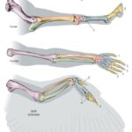
Alt: Illustration of sheep brain anatomy, highlighting key structures like the cerebrum, cerebellum, and brainstem in a detailed anatomical diagram.
References
-
Felix, R.; Gaser, C.; Hildebrandt, T.; Scheffler, F.; Wree, A.; Henkel, A. Stereotaxic brain atlas for the pig suitable for magnetic resonance imaging. J. Neurosci. Methods 2010, 194, 62–68.
-
Ella, S.R.; Al-Rubaie, S.; Ghoreishizadeh, S.S.; Nasrallah, F.; Harding, A.J.; Gamage, K.K.; MacLeod, A.; Rosin, P.L.; Tavakoli, T.; Pitelka, V.; et al. Generation of a high-resolution in vivo MRI stereotaxic atlas of the sheep brain. PLoS One 2020, 15, e0232834.
-
Paxinos, G.; Franklin, K.B.J. The Mouse Brain in Stereotaxic Coordinates, Compact Third Edition; Academic Press: San Diego, CA, USA, 2007.
-
Saleem, K.S.; Logothetis, N.K. A combined MRI and Stereotaxy Atlas of the Macaque Brain; Academic Press: San Diego, CA, USA, 2012.
-
McDonald, J.W.; Augustine, J.R.; Sotnikov, V.A.; Smith, D.W.; Toga, A.W. A probabilistic atlas of the cat brain. Neuroimage 2008, 42, 1401–1407.
-
Swanson, L.W. Brain Maps: Structure of the Rat Brain, 3rd ed.; Elsevier: Amsterdam, The Netherlands, 2004.
-
Paxinos, G.; Watson, C. The Rat Brain in Stereotaxic Coordinates, 6th ed.; Academic Press: San Diego, CA, USA, 2007.
-
Dittrich, H.; Gaser, C.; Wree, A.; Meyer, R.; Driesnack, M.; Henkel, A. A three-dimensional digital atlas of the beagle brain in stereotaxic coordinates. BMC Neurosci. 2009, 10, 135.
-
Frontera, B.; Garosi, L.; McConnell, J.; De Risio, L. Development of a canine brain magnetic resonance imaging template. Vet. Radiol. Ultrasound 2013, 54, 515–523.
-
Mai, J.K.; Paxinos, G.; Ashwell, K.W.B. The Human Nervous System, 3rd ed.; Academic Press: San Diego, CA, USA, 2016.
-
Dell’Anna, M.M.; Driver, C.J.; McGonnell, I.M.; Volk, H.A. Similarities and differences between the architecture of white matter in the human and sheep brain: A diffusion tensor imaging study. Brain Struct. Funct. 2016, 221, 2895–2915.
-
Gonzalez, J.A.; Forero, M.; Insausti, R. Cytoarchitectonic organization of the sheep cerebral cortex. Brain. Struct. Funct. 2012, 217, 261–280.
-
Stolp, H.B.; Scahill, V.L.; Rishniw, M.; McGonnell, I.M.; Driver, C.J. Use of the sheep as a model for studying brain development and disorders. ILAR J. 2015, 56, 283–297.
-
Workman, J.L.; Tigno, X.T.; Allen, A.M.; Stowe, A.M.; Caralopoulos, I.N. Sheep as a translational large animal model of human neurological disorders. Front. Neurol. 2013, 4, 10.
-
Banks, W.J. Applied Veterinary Histology, 3rd ed.; Mosby: St. Louis, MO, USA, 1993.
-
Fletcher, T.F.; Dellmann, H.D. Textbook of Veterinary Histology, 2nd ed.; Lea & Febiger: Philadelphia, PA, USA, 1991.
-
Rogers, C.S.; VandePol, K.J.; McAllister, T.A.; Napper, B.A.; Olson, M.R.; Stokol, T.; Baird, J.D.; Maysinger, D.; Muthuswamy, S.K.; Bienzle, D.; et al. Preclinical models of primary brain cancer: A comparative analysis of human, canine, and porcine gliomas. ILAR J. 2017, 58, 46–61.
-
Snyder, R.L.; Cheeke, P.R.; Schmitz, J.A. The Nervous System; Oregon State University: Corvallis, OR, USA, 2018; Available online: https://open.oregonstate.education/nervoussystem/ (accessed on May 2, 2023).
-
Knowlton, E.; Witonsky, J.; Capra, B.; Weiss, L.; Garlinghouse, M.; Marini, J.; Spurlock, M.; Druzgal, T.J.; Khalil, A.; Brock, M.; et al. Sonothrombolysis of experimental carotid artery thrombosis in sheep using transcranial focused ultrasound. J. Transl. Med. 2020, 18, 124.
-
Nowak, M.P.; Morrison, J.H.; Hof, P.R. Cortical microcircuits: Integrative substrates for cognition. Trends Neurosci. 2003, 26, 30–39.
-
Haight, J.R.; Neylon, A.J.;影響, S.M. The organization of neocortex in the sheep (Ovis aries): I. motor and somatosensory cortex. Somatosens. Mot. Res. 1987, 5, 255–278.
-
Gonzalez, J.A.; Weruaga, E.; Argandoña, E.G.; Carretero, N.; Garcia-Amado, M.; Insausti, R. Laminar distribution of calretinin-immunoreactive neurons in the sheep cerebral cortex. J. Chem. Neuroanat. 2009, 37, 1–12.
-
Marin, O. Interneuron dysfunction in psychiatric disorders. Nat. Rev. Neurosci. 2012, 13, 107–120.
-
Getty, R. Sisson and Grossman’s the Anatomy of the Domestic Animals; W.B. Saunders: Philadelphia, PA, USA, 1975; Volume 1–2.
-
Gasser, R.B.; Sprenger, N.; Gaschen, F.P.; Seewald, W.; Sigrist, B.; Francey, T.; Hilbe, M.; Howard, J.; Torgerson, P.R. Determination of reference intervals for selected clinical biochemical analytes in llamas (Lama glama). Vet. Clin. Pathol. 2004, 33, 219–224.
-
Onar, V.; Gunduz, S.; Ocal, M.K.; Yilmaz, S.; Agca, C.A. Anatomical and computed tomographic study of the orbit in sheep. Anat. Histol. Embryol. 2011, 40, 461–467.
-
Dell’Anna, M.M.; Driver, C.J.; Volk, H.A. Evaluation of white matter microstructure in sheep using diffusion tensor imaging. Vet. Radiol. Ultrasound 2014, 55, 202–212.
-
McGonnell, I.M.; Driver, C.J.; Brodbelt, D.C.; Volk, H.A. Magnetic resonance imaging of the normal ovine brain. Vet. Radiol. Ultrasound 2010, 51, 620–629.
-
Dyce, K.M.; Sack, W.O.; Wensing, C.J.G. Textbook of Veterinary Anatomy; W.B. Saunders: Philadelphia, PA, USA, 1987.
-
Kaplan, R.S.; Gonzalez, J.A.; Insausti, R.; Gomez-Nicola, D.; Stolp, H.B. Differential expression of doublecortin and polysialylated neural cell adhesion molecule reveals variations in the subventricular zone of the sheep brain. J. Comp. Neurol. 2011, 519, 3231–3247.
-
Albin, R.L.; Patnaik, M.; Bao, F.; Clayburgh, D.; Kotagal, V.; Bockenstedt, K.; Westbrook, L.; McKeever, P.; Anderson, K.D.; Young, A.B.; et al. The cellular and neurochemical anatomy of the human substantia nigra. J. Comp. Neurol. 1992, 326, 567–581.
-
DeCamp, C.E.; Doherty, T.; Lu, Y.; Kearney, M.T.; Johnston, S.A.; Schwarz, P.D. Radiographic anatomy of the sheep spine. Vet. Surg. 2010, 39, 515–520.
-
Khan, S.; Gill, P.; Schneider, I.; Thomson, C.E.; Hanrahan, B.M.; Singh, A.; Singh, G.; Singh, N. Sciatic nerve block in sheep: A cadaveric study to determine the optimal injection site using methylene blue dye. N. Z. Vet. J. 2023, 71, 146–152.
-
Arendt, J.; Skene, D.J. Melatonin as a marker for circadian rhythmicity. Chronobiol. Int. 2005, 22, 19–34.
-
Richard, P. Atlas Stereotaxique du Cerveau de Brebis; I.N.R.A.: Paris, France, 1967.
-
Heath, T.J. Outline of sheep anatomy. 1980; Available online: https://hdl.handle.net/102.100.100/308738?index=1 (accessed on May 2, 2023).
-
Maracacci, A. Studii Comparativi Sulla Fisiologia Degli Organi Nervosi Encefalo Midollo Spinale Midollo Allungato; G. Antonelli: Venezia, Italy, 1877.
-
Dusser de Barenne, J.G. Experimental researches on sensory localization in the cerebral cortex of the monkey (Macacus rhesus). Proc. R. Soc. Lond. Ser. B-Biol. Sci. 1924, 96, 272–291.
-
Bailey, P.; Bremer, F. A sensory cortical representation of the vagus nerve with a note on the effects of low blood pressure on the cortical electrogram. J. Neurophysiol. 1938, 1, 405–412.
-
McKenzie, J.S.; Smith, D.H. Stereotaxic coordinates and variation in the sheep brain. J. Physiol. 1973, 233, 72P–73P.
-
Opdam, H.I.M.; Van Rijen, P.C.; Van Nieuwenhuizen, O.; Backes, W.H.; Nicolay, K.; Vles, J.S.H. Sheep model of focal epilepsy: Concurrent EEG and functional MRI. Epilepsia 2002, 43, 1079–1087.
-
Badea, A.; Ali, A.; Johnson, G.A. In vivo atlas of the C57BL/6J mouse brain. Neuroimage 2007, 36, 1308–1316.
-
Collins, M.J.; Harding, A.J.; MacLeod, A.; Rosin, P.L.; Cowan, R.; Johnston, L.A. Development of an MRI-based atlas of the normal sheep brain and its application to define the spatial distribution of lesions in a transmissible spongiform encephalopathy. BMC Vet. Res. 2015, 11, 31.
-
Nitzsche, L.; Puster, P.; Böhme, R.; Planert, M.; Fischer, T.; Rösch, S.; Ondřejčka, R.; Gaser, C.; Boltze, J. An MRI-based ovine brain template and tissue probability maps. Brain Struct. Funct. 2015, 220, 2533–2542.
-
Mainau, E.; Muñoz, D.; Velarde, A.; Dalmau, A.; Temple, D.; Manteca, X. Assessment of brain and pituitary volume in lambs with or without tail docking using magnetic resonance imaging. Res. Vet. Sci. 2012, 93, 826–829.
-
Nowland, S.; Lewis, C.; Lawson, J.; Joers, V.; Bilkey, D.; Roxburgh, R. Frameless MRI guided stereotactic access to the ovine brainstem. J. Neurosci. Methods 2014, 222, 233–239.
-
Russell, T.A.; Gill, J.; Grieve, S.M.; van der Knapp, M.S.; Leventer, R.J.; Williams, R.E.; Fahey, M.C.; Bellgard, M.; Tam, P.P.L.; Bye