Are you trying to understand the fundamental differences between mitosis and meiosis? At COMPARE.EDU.VN, we provide a comprehensive breakdown of these crucial cell division processes, including a focus on chromosome behavior, key stages, and overall function. Understanding these variations is essential for grasping genetics and heredity.
1. What Are the Key Differences Between Mitosis and Meiosis?
Mitosis and meiosis are both types of cell division, but they serve distinct purposes and have key differences. Mitosis produces two genetically identical daughter cells, while meiosis produces four genetically unique daughter cells with half the number of chromosomes.
Mitosis is essential for growth, repair, and asexual reproduction. Meiosis, on the other hand, is specialized for sexual reproduction, creating gametes (sperm and egg cells) that combine to form a new organism. Let’s delve deeper into the specifics:
1.1. Purpose
- Mitosis: For cell growth, repair, and asexual reproduction. It ensures that each new cell receives an identical copy of the parent cell’s chromosomes.
- Meiosis: For sexual reproduction. It produces gametes with half the number of chromosomes, ensuring genetic diversity in offspring.
1.2. Number of Divisions
- Mitosis: Involves one round of cell division.
- Meiosis: Involves two rounds of cell division (Meiosis I and Meiosis II).
1.3. Chromosome Number
- Mitosis: The chromosome number remains the same in daughter cells (diploid to diploid).
- Meiosis: The chromosome number is halved in daughter cells (diploid to haploid).
1.4. Genetic Variation
- Mitosis: Daughter cells are genetically identical to the parent cell.
- Meiosis: Daughter cells are genetically unique due to crossing over and independent assortment.
1.5. Pairing of Homologous Chromosomes
- Mitosis: Homologous chromosomes do not pair up.
- Meiosis: Homologous chromosomes pair up during Prophase I (synapsis).
1.6. Crossing Over
- Mitosis: Crossing over does not occur.
- Meiosis: Crossing over occurs during Prophase I, leading to genetic recombination.
1.7. Resulting Daughter Cells
- Mitosis: Two diploid daughter cells.
- Meiosis: Four haploid daughter cells.
2. What Are the Stages of Mitosis and Meiosis?
Mitosis and meiosis share some similar stages but differ significantly in how these stages are executed, particularly in meiosis I.
2.1. Stages of Mitosis
Mitosis consists of four main stages:
- Prophase: Chromosomes condense, and the nuclear envelope breaks down. The spindle fibers begin to form.
- Metaphase: Chromosomes align at the metaphase plate (the center of the cell).
- Anaphase: Sister chromatids separate and move to opposite poles of the cell.
- Telophase: Chromosomes arrive at the poles, and the nuclear envelope reforms. Cytokinesis (cell division) usually occurs, resulting in two identical daughter cells.
2.2. Stages of Meiosis
Meiosis consists of two divisions: Meiosis I and Meiosis II.
2.2.1. Meiosis I
-
Prophase I: This is the longest and most complex phase of meiosis. It is further divided into:
- Leptotene: Chromosomes begin to condense.
- Zygotene: Homologous chromosomes pair up (synapsis).
- Pachytene: Crossing over occurs between homologous chromosomes.
- Diplotene: Homologous chromosomes begin to separate but remain attached at chiasmata (points where crossing over occurred).
- Diakinesis: Chromosomes are fully condensed, and the nuclear envelope breaks down.
-
Metaphase I: Homologous chromosome pairs align at the metaphase plate.
-
Anaphase I: Homologous chromosomes separate and move to opposite poles. Sister chromatids remain together.
-
Telophase I: Chromosomes arrive at the poles, and the cell divides. Each daughter cell now has a haploid set of chromosomes.
2.2.2. Meiosis II
Meiosis II is very similar to mitosis.
- Prophase II: Chromosomes condense, and the nuclear envelope breaks down (if it reformed during Telophase I).
- Metaphase II: Chromosomes align at the metaphase plate.
- Anaphase II: Sister chromatids separate and move to opposite poles.
- Telophase II: Chromosomes arrive at the poles, and the nuclear envelope reforms. Cytokinesis occurs, resulting in four haploid daughter cells.
3. How Do Chromosomes Behave Differently in Mitosis and Meiosis?
Chromosome behavior is a critical factor differentiating mitosis and meiosis.
3.1. Mitosis
- Duplication: Chromosomes duplicate during the S phase of the cell cycle, resulting in sister chromatids.
- Alignment: Sister chromatids align independently at the metaphase plate.
- Segregation: During anaphase, sister chromatids separate, ensuring each daughter cell receives an identical set of chromosomes.
- Cohesion: Sister chromatids are connected by the cohesin complex, which is cleaved during anaphase to allow separation.
3.2. Meiosis
- Duplication: Chromosomes duplicate during the S phase, as in mitosis.
- Pairing: Homologous chromosomes pair up during Prophase I in a process called synapsis.
- Crossing Over: Genetic material is exchanged between homologous chromosomes during Prophase I, leading to genetic recombination.
- Alignment: Homologous chromosome pairs align at the metaphase plate in Metaphase I.
- Segregation in Meiosis I: Homologous chromosomes separate during Anaphase I, but sister chromatids remain together.
- Segregation in Meiosis II: Sister chromatids separate during Anaphase II, similar to mitosis.
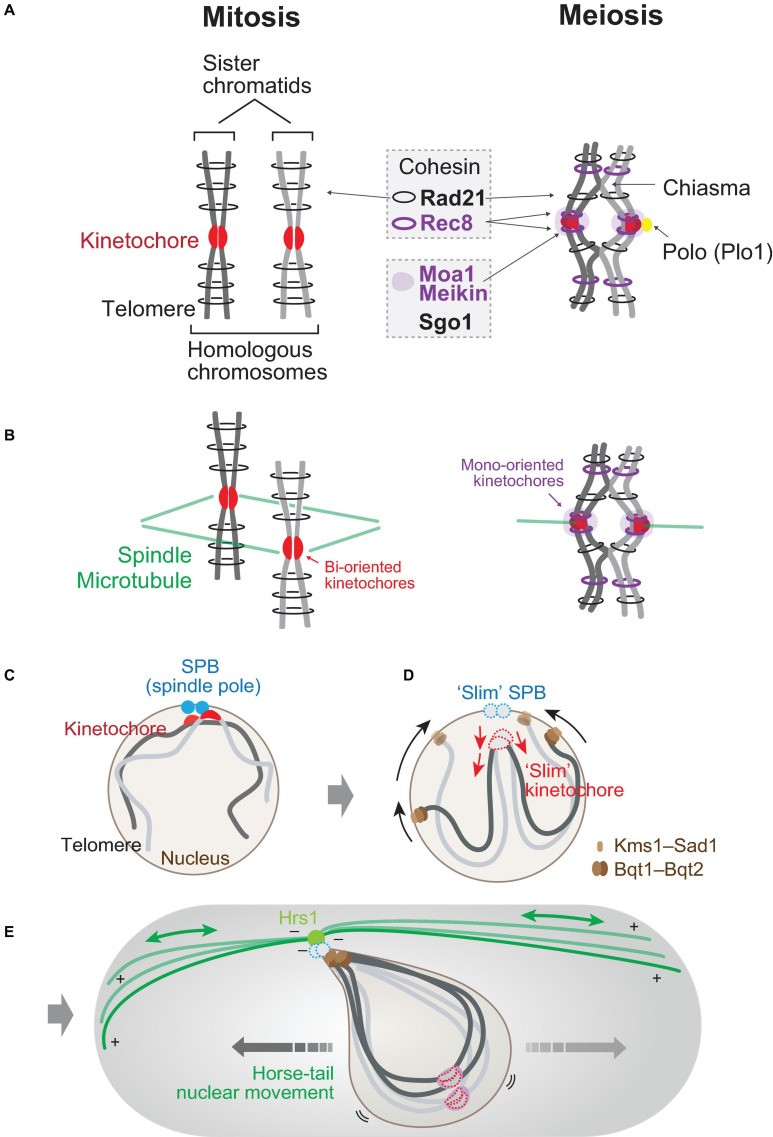
- Cohesion: Meiotic cohesin (Rec8) maintains cohesion between sister chromatids and homologous chromosomes. Rec8 is protected at the centromere during Anaphase I by shugoshin, ensuring sister chromatids remain together until Anaphase II.
4. What Is the Significance of Genetic Variation in Meiosis?
Genetic variation, a hallmark of meiosis, is crucial for evolution and adaptation. This variation arises through two primary mechanisms: crossing over and independent assortment.
4.1. Crossing Over
- Process: Exchange of genetic material between homologous chromosomes during Prophase I.
- Result: Creates new combinations of genes on each chromosome, increasing genetic diversity.
4.2. Independent Assortment
- Process: Random alignment of homologous chromosome pairs at the metaphase plate during Metaphase I.
- Result: Each daughter cell receives a different combination of maternal and paternal chromosomes, further enhancing genetic diversity.
4.3. Implications
- Evolution: Genetic variation provides the raw material for natural selection, allowing populations to adapt to changing environments.
- Disease Resistance: Genetic diversity increases the likelihood that some individuals will have traits that confer resistance to diseases.
- Agriculture: Genetic variation is essential for breeding new crop varieties with improved traits.
5. How Are Kinetochores and Microtubules Involved in Mitosis and Meiosis?
Kinetochores and microtubules play essential roles in chromosome segregation during both mitosis and meiosis.
5.1. Mitosis
- Kinetochores: Protein structures assembled on the centromeres of chromosomes.
- Microtubules: Spindle fibers that attach to kinetochores and pull chromosomes apart.
- Attachment: Sister kinetochores attach to microtubules from opposite poles of the cell (bi-orientation).
- Segregation: Microtubules shorten, pulling sister chromatids to opposite poles.
5.2. Meiosis I
- Kinetochores: As in mitosis.
- Microtubules: As in mitosis.
- Attachment: Sister kinetochores attach to microtubules from the same pole of the cell (mono-orientation) in Meiosis I.
- Segregation: Microtubules shorten, pulling homologous chromosomes to opposite poles. Sister chromatids remain together.
5.3. Meiosis II
- Kinetochores: As in mitosis.
- Microtubules: As in mitosis.
- Attachment: Sister kinetochores attach to microtubules from opposite poles of the cell, similar to mitosis.
- Segregation: Microtubules shorten, pulling sister chromatids to opposite poles.
6. What Are the Consequences of Errors in Mitosis and Meiosis?
Errors in mitosis and meiosis can have significant consequences, ranging from developmental abnormalities to cancer.
6.1. Mitosis Errors
- Non-disjunction: Failure of sister chromatids to separate properly.
- Aneuploidy: Resulting cells have an abnormal number of chromosomes.
- Cancer: Errors in mitosis can lead to uncontrolled cell division and tumor formation.
6.2. Meiosis Errors
- Non-disjunction: Failure of homologous chromosomes (Meiosis I) or sister chromatids (Meiosis II) to separate properly.
- Aneuploidy in Gametes: Gametes have an abnormal number of chromosomes.
- Genetic Disorders: Fertilization with an aneuploid gamete can lead to genetic disorders such as Down syndrome (trisomy 21).
- Miscarriage: Many aneuploid embryos are not viable and result in miscarriage.
7. What Are the Key Proteins Involved in Mitosis and Meiosis?
Several key proteins regulate the processes of mitosis and meiosis.
7.1. Mitosis Proteins
- Cyclin-Dependent Kinases (CDKs): Regulate the cell cycle progression.
- Cohesin: Holds sister chromatids together.
- Kinesin and Dynein: Motor proteins involved in spindle formation and chromosome movement.
- Spindle Assembly Checkpoint (SAC) Proteins: Ensure proper chromosome attachment to microtubules.
7.2. Meiosis Proteins
- Rec8: Meiosis-specific cohesin.
- Moa1 (Meikin): Regulates kinetochore mono-orientation in Meiosis I.
- Shugoshin: Protects Rec8 at the centromere during Anaphase I.
- Polo-like Kinase (Plo1): Regulates kinetochore-microtubule interactions.
- MutS Homolog 4 (MSH4) and MutL Homolog 1 (MLH1): Facilitate meiotic recombination.
8. How Does Sexual Differentiation Affect Chromosome Arrangement?
Sexual differentiation profoundly affects chromosome arrangement, especially in preparation for meiosis.
8.1. Mitotic Cell Cycles
- Interphase: Centromeres of all chromosomes cluster near spindle pole bodies (SPBs).
- Rabl Orientation: This arrangement facilitates easy connection between microtubules and kinetochores upon mitotic entry.
8.2. Sexual Differentiation in Meiosis
- Response to Mating Pheromone: Telomeres cluster, and centromeres dissociate from SPBs.
- Bouquet Arrangement: Telomeres are clustered at SPBs, while centromeres are located far from SPBs.
- Horse-Tail Nuclear Movement: Microtubule-driven movement aligns chromosomes for pairing and recombination.
- LINC Complex: Telomere proteins (Bqt1, Bqt2) bind to nuclear membrane proteins (Sad1, Kms1/2) to bring telomeres to SPBs.
9. How Does Entry into Meiosis I Alter Chromosome Position?
Chromosome positioning is significantly altered upon entry into meiosis I to facilitate the unique events of this division.
9.1. Mitotic Entry
- Kinetochore Proximity: Kinetochores are located close to SPBs.
- Microtubule Nucleation: Microtubules nucleate from SPBs, easily connecting with kinetochores.
9.2. Meiotic Entry
- Upside-Down Positioning: Centromeres dissociate, and telomeres cluster, inverting chromosome arrangement.
- Radial Array of Microtubules: Extensive radial microtubules nucleate from SPBs, capturing scattered kinetochores.
- Kinetochore Retrieval: Microtubules shrink to retrieve attached kinetochores toward SPBs.
- Alp7 (TACC) Localization: Alp7 localizes to kinetochores, promoting capture by radial microtubules.
10. How Is the Spindle Assembled Differently in Mitosis and Meiosis?
The assembly of the spindle, which is crucial for chromosome segregation, differs significantly between mitosis and meiosis.
10.1. Mitotic Spindle Assembly
- Microtubule Elongation: Microtubules elongate from two SPBs.
- SPB Separation: SPBs separate outward due to the interaction of microtubules.
- Kinesin Motor Proteins: Kinesin-5 (Cut7) facilitates SPB separation, while kinesin-14 (Pkl1, Klp2) generates inward forces.
- Chromosome-Mediated Outward Force: Sister chromatids mediate pole-to-pole connection through kinetochore-microtubule attachment.
10.2. Meiotic Spindle Assembly
- Homologous Kinetochore Connection: Loosened connection between homologous kinetochores.
- Mono-Orientation: Sister chromatids are mono-oriented, resulting in weaker kinetochore-mediated outward force.
- Cut7 Dependence: Meiosis I is more dependent on Cut7-mediated force for SPB separation.
11. What Is the Role of Anaphase Promoting Complex/Cyclosome (APC/C) in Meiosis?
The APC/C plays a crucial role in regulating the progression through meiosis by controlling the degradation of key regulatory proteins.
11.1. APC/C Activation
- Coactivators: Cdc20 (Slp1) and Fzr1 activate APC/C.
- Meiosis I: Slp1 is the main coactivator for anaphase I onset.
- Meiosis II: Fzr1 assists in APC/C activation for anaphase II onset and completion.
11.2. APC/C Inhibitor: Mes1
- Function: Inhibits APC/C to maintain CDK activity, allowing cells to enter anaphase I and restart meiosis II.
- Regulation: Binds and inhibits Fzr1 and Slp1, preventing premature APC/C activation.
11.3. Consequences of APC/C Dysregulation
- mes1Δ Mutant: APC/C is prematurely activated, terminating meiosis early.
- fzr1Δ Mutant: Meiosis III occurs due to insufficient CDK repression after anaphase II.
12. How Does Meiosis II Differ From Mitosis?
While meiosis II shares similarities with mitosis, it also exhibits unique features, particularly in the context of gametogenesis.
12.1. Similarities
- Equational Division: Sister chromatids separate, similar to mitosis.
- Chromosome Segregation: The basic mechanisms of chromosome segregation are analogous to mitosis.
12.2. Differences
- Forespore Membrane Formation: SPBs are modified to assemble the forespore membrane, which surrounds the nucleus.
- Virtual Nuclear Envelope Breakdown (vNEBD): The nuclear envelope becomes permeable, allowing nucleoplasmic proteins to disperse.
- Dispensable Interpolar Microtubules: The forespore membrane can serve as an interpolar structure, making interpolar microtubules dispensable in meiosis II.
13. What Are Some Frequently Asked Questions About Mitosis and Meiosis?
Q1: What is the main purpose of mitosis?
Mitosis is primarily for cell growth, repair, and asexual reproduction, ensuring each new cell receives an identical copy of the parent cell’s chromosomes.
Q2: What is the main purpose of meiosis?
Meiosis is specialized for sexual reproduction, creating genetically diverse gametes (sperm and egg cells) with half the number of chromosomes.
Q3: How many daughter cells are produced in mitosis and meiosis?
Mitosis produces two diploid daughter cells, while meiosis produces four haploid daughter cells.
Q4: What is crossing over, and when does it occur?
Crossing over is the exchange of genetic material between homologous chromosomes during Prophase I of meiosis, leading to genetic recombination.
Q5: What is non-disjunction, and what are its consequences?
Non-disjunction is the failure of chromosomes to separate properly during cell division. It can lead to aneuploidy, resulting in cells with an abnormal number of chromosomes and potentially causing genetic disorders or cancer.
Q6: What is the role of kinetochores in mitosis and meiosis?
Kinetochores are protein structures on centromeres where microtubules attach, facilitating chromosome movement and segregation during both mitosis and meiosis.
Q7: What is the significance of genetic variation in meiosis?
Genetic variation, created through crossing over and independent assortment, is crucial for evolution, disease resistance, and the adaptation of populations to changing environments.
Q8: How does the spindle assembly differ in mitosis and meiosis?
In mitosis, sister chromatids align independently, while in meiosis I, homologous chromosomes pair and align together. This difference affects the forces and mechanisms involved in spindle pole separation.
Q9: What are some key proteins involved in mitosis and meiosis?
Key proteins include CDKs, cohesin, kinesin, dynein, Rec8, Moa1 (Meikin), shugoshin, and Polo-like Kinase (Plo1). Each plays a specific role in regulating cell cycle progression and chromosome segregation.
Q10: How does sexual differentiation affect chromosome arrangement in meiosis?
Sexual differentiation leads to telomere clustering and centromere dissociation from spindle pole bodies, forming the “bouquet” arrangement that promotes pairing and meiotic recombination of homologous chromosomes.
Mitosis and meiosis are vital processes with distinct purposes and outcomes. Understanding their nuances is essential for grasping genetics, heredity, and the broader scope of life sciences. Whether you’re a student, a consumer, or a professional, COMPARE.EDU.VN provides you with the knowledge you need to make informed decisions.
COMPARE.EDU.VN: Your Partner in Informed Decisions
At COMPARE.EDU.VN, we’re dedicated to providing detailed, objective comparisons to help you make the best choices, whether you’re comparing academic concepts, consumer products, or professional methodologies. Our goal is to simplify complex information and empower you to make confident decisions.
Navigating the complexities of cellular biology, or any other subject, can be daunting. But with the right resources, you can clarify your understanding and make informed decisions.
Ready to Explore More?
Don’t let the complexities of mitosis and meiosis, or any other comparison, hold you back. Visit COMPARE.EDU.VN today to discover comprehensive comparisons that empower your decisions.
Address: 333 Comparison Plaza, Choice City, CA 90210, United States
WhatsApp: +1 (626) 555-9090
Website: COMPARE.EDU.VN
Visit compare.edu.vn to unlock a world of knowledge and make comparisons easier than ever before.