A comparative view of sperm ultrastructure, particularly as pioneered by Don Fawcett, is indeed important for understanding reproductive biology, diagnosing infertility, and developing new contraceptive methods, all of which COMPARE.EDU.VN aims to clarify. Fawcett’s work laid the foundation for understanding sperm cell biology and its implications for fertility and reproductive health. Exploring seminal cytology and sperm morphology enhances our comprehension of these critical factors.
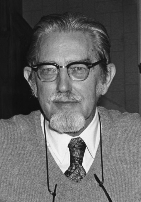
1. Who Was Don Fawcett and Why Is His Work on Sperm Ultrastructure Important?
Don Fawcett (1917-2009) was a highly influential anatomist and cell biologist whose research significantly advanced our understanding of cell structure and function, especially in reproductive biology. His comparative anatomical analysis of sperm cell ultrastructure provided foundational knowledge in the field.
1.1 Fawcett’s Early Life and Education
Born in Iowa, Don Fawcett’s academic journey began at the prestigious Boston Latin School. He pursued higher education at Harvard College and Harvard Medical School, where he was deeply influenced by George B. Wislocki, the chairman of the Department of Anatomy.
1.2 Contributions to Cell Biology
Fawcett’s pioneering work in electron microscopy in the 1950s revolutionized cell biology. His early studies included detailed descriptions of cilia and sperm flagella. These foundational works established him as a leading figure in the field.
1.3 Research on Male Reproductive System
Fawcett’s collaboration with M.H. Burgos in 1955 marked the beginning of his extensive research on the male reproductive system. Their work on the fine structure and development of cat spermatids was followed by significant publications on spermatid differentiation in toads.
1.4 Landmark Contribution to Reproductive Biology
In 1965, Fawcett published a landmark paper describing the guinea pig sperm during and after differentiation. This work is considered a seminal contribution to reproductive biology and solidified his reputation as a leading expert in the field.
1.5 Collaborative Research and Discoveries
During the late 1960s through the early 1980s, Fawcett collaborated with numerous faculty members, fellows, students, and international scientists. These collaborations led to discoveries such as the junctional specializations of Sertoli cells, which form the structural basis of the blood-testis barrier.
2. What is Sperm Ultrastructure?
Sperm ultrastructure refers to the detailed microscopic anatomy of sperm cells, including the various components and organelles that make up the sperm. This includes the head, midpiece, and tail, each with distinct structures essential for sperm function.
2.1 Components of Sperm Ultrastructure
The basic components of sperm ultrastructure include the head (containing the nucleus and acrosome), the midpiece (containing mitochondria), and the tail (flagellum). Each component has a specific function critical for fertilization.
2.1.1 Sperm Head
The sperm head contains the nucleus, which carries the genetic material, and the acrosome, a cap-like structure containing enzymes necessary for penetrating the egg.
2.1.2 Midpiece
The midpiece is packed with mitochondria, which provide the energy required for the sperm to swim and reach the egg.
2.1.3 Tail (Flagellum)
The tail, or flagellum, is responsible for the sperm’s motility. Its whip-like movements propel the sperm through the female reproductive tract.
2.2 Techniques for Studying Sperm Ultrastructure
Electron microscopy is the primary technique used to study sperm ultrastructure. This technique provides high-resolution images of the sperm’s internal components, allowing for detailed analysis.
2.2.1 Transmission Electron Microscopy (TEM)
TEM is used to visualize the internal structure of sperm cells. It allows researchers to examine the arrangement of organelles and other components at a very high magnification.
2.2.2 Scanning Electron Microscopy (SEM)
SEM is used to examine the surface features of sperm cells. It provides a three-dimensional view of the sperm’s external morphology.
3. Why is a Comparative View of Sperm Ultrastructure Important?
A comparative view of sperm ultrastructure allows for a better understanding of the variations in sperm morphology and function across different species. This knowledge is essential for addressing issues related to fertility and reproduction.
3.1 Understanding Infertility
Variations in sperm ultrastructure can affect sperm motility, viability, and the ability to fertilize an egg. Identifying these abnormalities is crucial for diagnosing and treating infertility.
3.1.1 Sperm Morphology and Fertility
Abnormal sperm morphology can impair the sperm’s ability to swim and penetrate the egg. A comparative analysis helps in identifying these abnormalities.
3.1.2 Diagnosing Male Infertility
Sperm ultrastructure analysis is a valuable tool for diagnosing male infertility. It can reveal defects that are not visible with standard semen analysis techniques.
3.2 Reproductive Biology
Comparing sperm ultrastructure across species provides insights into the evolution of reproductive strategies. It helps in understanding the adaptations that enable successful fertilization in different environments.
3.2.1 Evolution of Sperm Morphology
Sperm morphology varies significantly across species, reflecting adaptations to different reproductive environments.
3.2.2 Comparative Anatomy of Sperm
A comparative view of sperm anatomy reveals how structural differences relate to functional variations in fertilization.
3.3 Developing Contraceptive Methods
Understanding sperm ultrastructure is essential for developing new contraceptive methods. Targeting specific components of sperm cells can lead to more effective and safer contraceptives.
3.3.1 Targeting Sperm Components
Contraceptive methods can be designed to target specific components of sperm cells, such as the acrosome or flagellum, to prevent fertilization.
3.3.2 Innovative Contraceptive Strategies
Advanced research in sperm ultrastructure can lead to innovative contraceptive strategies that are more effective and have fewer side effects.
4. What Were Don Fawcett’s Key Discoveries in Sperm Ultrastructure?
Don Fawcett made several key discoveries in sperm ultrastructure, including detailed descriptions of the acrosome, flagellum, and Sertoli cells. His work provided critical insights into the structure and function of these components.
4.1 Acrosome Structure and Function
Fawcett’s work on the acrosome revealed its complex structure and the role of its enzymes in penetrating the egg.
4.1.1 Acrosomal Enzymes
He identified the enzymes within the acrosome that are essential for breaking down the outer layers of the egg, allowing the sperm to fuse with the egg membrane.
4.1.2 Acrosome Reaction
Fawcett’s research contributed to understanding the acrosome reaction, the process by which the acrosome releases its enzymes to facilitate fertilization.
4.2 Flagellum Structure and Motility
Fawcett’s descriptions of the flagellum detailed its intricate structure and how it generates the force needed for sperm motility.
4.2.1 Flagellar Components
He identified the various components of the flagellum, including the axoneme and outer dense fibers, and explained their roles in motility.
4.2.2 Sperm Motility Mechanisms
Fawcett’s work elucidated the mechanisms by which the flagellum generates movement, enabling the sperm to swim towards the egg.
4.3 Sertoli Cells and Blood-Testis Barrier
Fawcett’s research on Sertoli cells revealed their crucial role in supporting spermatogenesis and forming the blood-testis barrier.
4.3.1 Sertoli Cell Function
He described how Sertoli cells provide nutrients and support to developing sperm cells within the seminiferous tubules.
4.3.2 Blood-Testis Barrier
Fawcett’s discovery of the junctional specializations of Sertoli cells explained the structural basis of the blood-testis barrier, which protects developing sperm from immune attack.
5. How Has Fawcett’s Work Influenced Current Research in Reproductive Biology?
Fawcett’s work has had a lasting impact on current research in reproductive biology, providing a foundation for advancements in fertility treatments, contraceptive development, and understanding genetic disorders.
5.1 Fertility Treatments
His research has informed current fertility treatments such as in vitro fertilization (IVF) and intracytoplasmic sperm injection (ICSI).
5.1.1 In Vitro Fertilization (IVF)
Fawcett’s detailed understanding of sperm ultrastructure has improved the selection of viable sperm for IVF procedures.
5.1.2 Intracytoplasmic Sperm Injection (ICSI)
ICSI involves injecting a single sperm directly into an egg. Fawcett’s research has helped refine the selection criteria for sperm used in ICSI.
5.2 Contraceptive Development
His discoveries have paved the way for developing new contraceptive methods that target specific sperm components.
5.2.1 Targeting Sperm Proteins
Current research focuses on identifying and targeting specific sperm proteins essential for fertilization, based on Fawcett’s foundational work.
5.2.2 Non-Hormonal Contraceptives
Researchers are exploring non-hormonal contraceptive options that disrupt sperm function, informed by Fawcett’s insights into sperm ultrastructure.
5.3 Understanding Genetic Disorders
His work has contributed to understanding genetic disorders that affect sperm structure and function, such as Kartagener syndrome.
5.3.1 Kartagener Syndrome
Kartagener syndrome is a genetic disorder that affects the structure of cilia and flagella, including the sperm tail. Fawcett’s research has helped elucidate the underlying mechanisms of this disorder.
5.3.2 Genetic Basis of Infertility
His work has contributed to understanding the genetic basis of infertility related to sperm defects, enabling better diagnosis and genetic counseling.
6. What is the Role of Electron Microscopy in Studying Sperm?
Electron microscopy plays a crucial role in studying sperm by providing high-resolution images of their internal and external structures. This technique is essential for identifying subtle abnormalities that cannot be detected with other methods.
6.1 High-Resolution Imaging
Electron microscopy allows for detailed visualization of sperm organelles and components, providing insights into their structure and function.
6.1.1 Detailed Visualization of Organelles
The high magnification capabilities of electron microscopy reveal the intricate details of sperm organelles, such as the acrosome, mitochondria, and flagellum.
6.1.2 Identification of Subtle Abnormalities
Electron microscopy can identify subtle abnormalities in sperm ultrastructure that may contribute to infertility.
6.2 Different Types of Electron Microscopy
Different types of electron microscopy, such as TEM and SEM, offer complementary information about sperm structure.
6.2.1 Transmission Electron Microscopy (TEM)
TEM is used to examine the internal structure of sperm cells, providing high-resolution images of organelles and other components.
6.2.2 Scanning Electron Microscopy (SEM)
SEM is used to examine the surface features of sperm cells, providing a three-dimensional view of their external morphology.
6.3 Applications in Fertility Research
Electron microscopy is widely used in fertility research to study sperm structure, identify defects, and evaluate the effects of treatments on sperm quality.
6.3.1 Assessing Sperm Quality
Electron microscopy is used to assess sperm quality by evaluating the integrity of sperm organelles and identifying structural abnormalities.
6.3.2 Evaluating Treatment Effects
Researchers use electron microscopy to evaluate the effects of various treatments, such as medications or lifestyle changes, on sperm structure and function.
 Sperm structure illustrated
7. How Does Sperm Ultrastructure Vary Across Species?
Sperm ultrastructure varies significantly across species, reflecting adaptations to different reproductive strategies and environments. Comparative studies provide insights into the evolution of sperm morphology and function.
7.1 Variations in Head Morphology
Sperm head morphology varies widely across species, with differences in shape, size, and acrosome structure.
7.1.1 Species-Specific Head Shapes
Different species have evolved unique sperm head shapes that are adapted to their specific reproductive environments.
7.1.2 Acrosome Size and Structure
The size and structure of the acrosome also vary across species, reflecting differences in the requirements for penetrating the egg.
7.2 Variations in Midpiece Structure
The midpiece structure, including the number and arrangement of mitochondria, varies across species depending on the energy requirements for sperm motility.
7.2.1 Mitochondrial Arrangement
The arrangement of mitochondria in the midpiece can differ significantly across species, affecting sperm energy production.
7.2.2 Energy Requirements
Species with longer sperm migration distances tend to have more mitochondria in the midpiece to provide the necessary energy.
7.3 Variations in Tail Structure
Sperm tail structure, including the length and arrangement of flagellar components, varies across species depending on the mode of fertilization.
7.3.1 Flagellar Length
The length of the flagellum can vary significantly across species, affecting sperm swimming speed and efficiency.
7.3.2 Flagellar Components
The arrangement and composition of flagellar components, such as the axoneme and outer dense fibers, can also vary across species.
8. What are Common Sperm Ultrastructural Defects?
Common sperm ultrastructural defects include abnormalities in the head, midpiece, and tail, which can impair sperm motility, viability, and fertilization ability.
8.1 Head Defects
Head defects can include abnormal shapes, acrosome abnormalities, and nuclear abnormalities, all of which can affect sperm function.
8.1.1 Abnormal Head Shapes
Irregular head shapes can impair sperm motility and the ability to penetrate the egg.
8.1.2 Acrosome Abnormalities
Defects in the acrosome can prevent the sperm from releasing the enzymes needed to penetrate the egg.
8.1.3 Nuclear Abnormalities
Abnormalities in the sperm nucleus can affect the genetic material carried by the sperm, leading to fertilization failures or developmental issues.
8.2 Midpiece Defects
Midpiece defects can include mitochondrial abnormalities and structural defects, which can impair sperm energy production and motility.
8.2.1 Mitochondrial Abnormalities
Defects in the mitochondria can reduce the energy available for sperm motility, affecting their ability to reach the egg.
8.2.2 Structural Defects
Structural defects in the midpiece can impair sperm motility and viability.
8.3 Tail Defects
Tail defects can include abnormal tail length, coiling, and structural defects, all of which can impair sperm motility.
8.3.1 Abnormal Tail Length
Sperm tails that are too short or too long can have reduced motility.
8.3.2 Tail Coiling
Coiled tails can impair sperm motility and the ability to swim towards the egg.
8.3.3 Structural Defects
Structural defects in the tail can affect sperm motility and viability.
9. How Can Sperm Ultrastructure Analysis Improve Fertility Outcomes?
Sperm ultrastructure analysis can improve fertility outcomes by providing detailed information about sperm quality, which can guide treatment decisions and improve the selection of viable sperm for assisted reproductive technologies.
9.1 Detailed Sperm Quality Assessment
Sperm ultrastructure analysis provides a more detailed assessment of sperm quality than standard semen analysis techniques.
9.1.1 Identifying Subtle Defects
Electron microscopy can identify subtle defects in sperm ultrastructure that may not be detected with other methods.
9.1.2 Guiding Treatment Decisions
The results of sperm ultrastructure analysis can help guide treatment decisions, such as choosing the most appropriate assisted reproductive technology.
9.2 Improved Sperm Selection
Sperm ultrastructure analysis can improve the selection of viable sperm for assisted reproductive technologies, such as IVF and ICSI.
9.2.1 Selecting Viable Sperm
By identifying and selecting sperm with normal ultrastructure, fertility specialists can improve the chances of successful fertilization.
9.2.2 Enhancing IVF Success Rates
Improved sperm selection can enhance IVF success rates by ensuring that only the highest quality sperm are used for fertilization.
9.3 Personalized Treatment Plans
Sperm ultrastructure analysis can inform the development of personalized treatment plans tailored to the specific needs of each patient.
9.3.1 Tailored Interventions
By understanding the specific sperm defects present in a patient’s sample, fertility specialists can develop tailored interventions to address these issues.
9.3.2 Optimizing Fertility Outcomes
Personalized treatment plans can optimize fertility outcomes by addressing the underlying causes of male infertility.
10. What are the Future Directions in Sperm Ultrastructure Research?
Future directions in sperm ultrastructure research include advancements in imaging techniques, genetic analysis, and the development of new treatments for male infertility.
10.1 Advancements in Imaging Techniques
New imaging techniques, such as super-resolution microscopy and cryo-electron microscopy, are providing even more detailed insights into sperm ultrastructure.
10.1.1 Super-Resolution Microscopy
Super-resolution microscopy allows for the visualization of sperm structures at an unprecedented level of detail.
10.1.2 Cryo-Electron Microscopy
Cryo-electron microscopy enables the study of sperm structures in their native state, without the need for staining or fixation.
10.2 Genetic Analysis of Sperm Defects
Genetic analysis is being used to identify the genes responsible for sperm ultrastructural defects, leading to better diagnosis and potential gene therapies.
10.2.1 Identifying Genetic Causes
Researchers are working to identify the genetic causes of sperm defects by analyzing the genomes of infertile men.
10.2.2 Potential Gene Therapies
The identification of genetic causes of sperm defects may lead to the development of gene therapies to correct these issues.
10.3 Development of New Treatments
New treatments are being developed to address sperm ultrastructural defects, including medications, lifestyle changes, and advanced assisted reproductive technologies.
10.3.1 Medications and Supplements
Researchers are investigating the potential of medications and supplements to improve sperm ultrastructure and function.
10.3.2 Lifestyle Changes
Lifestyle changes, such as diet and exercise, may also improve sperm quality and ultrastructure.
10.3.3 Advanced Assisted Reproductive Technologies
Advanced assisted reproductive technologies, such as ICSI with morphological sperm selection, are being developed to improve fertility outcomes for men with sperm ultrastructural defects.
Understanding sperm ultrastructure, as championed by Don Fawcett, is critical for advancements in reproductive biology, fertility treatments, and contraceptive development. By continuing to explore the intricacies of sperm cell biology, we can improve outcomes for individuals and couples facing infertility challenges. For comprehensive comparisons and detailed analyses, visit COMPARE.EDU.VN, your trusted resource for informed decision-making.
Are you struggling with making informed decisions? At COMPARE.EDU.VN, we provide detailed and objective comparisons to help you make the right choices. Contact us at 333 Comparison Plaza, Choice City, CA 90210, United States, or reach out via WhatsApp at +1 (626) 555-9090. Visit our website compare.edu.vn today to explore our comprehensive resources.
FAQ: Sperm Ultrastructure
1. What is sperm ultrastructure?
Sperm ultrastructure refers to the detailed microscopic anatomy of sperm cells, including the head, midpiece, and tail.
2. Why is a comparative view of sperm ultrastructure important?
It helps understand variations in sperm morphology and function across species, essential for addressing fertility and reproduction issues.
3. Who was Don Fawcett?
Don Fawcett was an influential anatomist and cell biologist whose research significantly advanced our understanding of cell structure and function, especially in reproductive biology.
4. What were Don Fawcett’s key discoveries in sperm ultrastructure?
He made key discoveries on the acrosome, flagellum, and Sertoli cells, providing critical insights into their structure and function.
5. How has Fawcett’s work influenced current research in reproductive biology?
His work has informed fertility treatments like IVF and ICSI, contraceptive development, and understanding genetic disorders affecting sperm.
6. What is the role of electron microscopy in studying sperm?
Electron microscopy provides high-resolution images of sperm’s internal and external structures, essential for identifying subtle abnormalities.
7. How does sperm ultrastructure vary across species?
It varies significantly, reflecting adaptations to different reproductive strategies and environments, with differences in head, midpiece, and tail morphology.
8. What are common sperm ultrastructural defects?
Common defects include abnormalities in the head, midpiece, and tail, which can impair sperm motility, viability, and fertilization ability.
9. How can sperm ultrastructure analysis improve fertility outcomes?
It provides detailed information about sperm quality, guiding treatment decisions and improving the selection of viable sperm for assisted reproductive technologies.
10. What are the future directions in sperm ultrastructure research?
Future directions include advancements in imaging techniques, genetic analysis, and the development of new treatments for male infertility.