A Comparative Study Of Deep Avian Foveas explores the structural and functional variations of the fovea, the region of highest visual acuity, in different bird species; COMPARE.EDU.VN is your premier destination for detailed comparisons. Understanding these differences sheds light on the evolutionary adaptations that enable birds to thrive in diverse ecological niches. Let’s embark on a comparative analysis, diving deep into avian vision, retinal morphology, and avian visual system.
1. What Is the Significance of Deep Avian Foveas?
Deep avian foveas are critical for high-acuity vision in birds, particularly diurnal species. They are specialized retinal structures characterized by a high density of photoreceptors, primarily cones, which enhances visual resolution and color perception.
The significance lies in:
- Enhanced Visual Acuity: Deep foveas allow birds to see fine details, crucial for hunting, foraging, and navigating complex environments.
- Predator Detection: Birds of prey rely on their deep foveas to spot prey from great distances with exceptional precision.
- Spatial Orientation: Accurate spatial vision is essential for flight and maneuvering through varied terrains.
- Color Vision: The high concentration of cones in the fovea enables vibrant color perception, aiding in mate selection and identifying food sources.
2. How Does the Structure of Deep Avian Foveas Compare Among Different Bird Species?
The structure of deep avian foveas varies significantly across different bird species, reflecting their diverse visual needs and ecological adaptations. Key structural differences include:
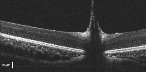
- Depth and Shape: Foveal depth and shape vary, with some species exhibiting deeper, steeper depressions compared to others. For example, diurnal birds of prey, such as hawks and eagles, possess very deep foveas, while other species have shallower depressions.
- Photoreceptor Density: The density of cones and rods within the fovea differs. Diurnal birds generally have a higher concentration of cones, while nocturnal birds may have a greater proportion of rods.
- Foveal Location: The position of the fovea on the retina can vary, with some birds having central foveas and others having temporal or nasal foveas. Some species even have multiple foveas to enhance their field of view and depth perception.
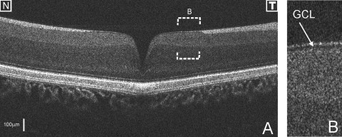
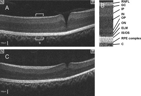
- Ganglion Cell Layer (GCL): The thickness and density of the GCL surrounding the fovea also differ, affecting signal processing and visual acuity.
- Retinal Nerve Fiber Layer (RNFL): The distribution and thickness of the RNFL near the fovea vary, influencing the overall visual field and sensitivity.
3. What Are the Functional Differences Between Deep and Shallow Avian Foveas?
Many diurnal birds, especially birds of prey, have two foveas: a deep (central or nasal) fovea and a shallow (temporal) fovea. These foveas serve different visual functions:
- Deep Fovea: Primarily used for high-resolution, binocular vision. It is essential for tasks requiring precise depth perception, such as judging distances when hunting or landing.
- Shallow Fovea: Often used for monocular vision and motion detection. It enhances the bird’s ability to detect movement in its peripheral vision, crucial for spotting predators or prey.
The comparative functional differences are summarized in the table below:
| Feature | Deep Fovea (Central/Nasal) | Shallow Fovea (Temporal) |
|---|---|---|
| Primary Function | High-resolution, binocular vision, precise depth perception | Monocular vision, motion detection |
| Use Case | Hunting, judging distances, detailed observation | Spotting predators or prey in peripheral vision |
| Photoreceptor Type | High cone density | Varied cone and rod density |
| Visual Acuity | High | Moderate |
| Field of View | Central | Peripheral |
4. How Do Diurnal and Nocturnal Birds Differ in Their Foveal Structure?
Diurnal and nocturnal birds exhibit distinct differences in their foveal structure, reflecting their adaptations to different light conditions:
- Diurnal Birds:
- High Cone Density: Deep foveas in diurnal birds are characterized by a high density of cones, allowing for excellent color vision and high visual acuity in bright light.
- Deep Foveal Pits: These birds often have pronounced foveal pits, which concentrate light onto the photoreceptors, enhancing visual resolution.
- Multiple Foveas: Some diurnal birds have multiple foveas, providing a wider field of high-acuity vision.
- Nocturnal Birds:
- High Rod Density: Foveas in nocturnal birds tend to have a higher density of rods, which are more sensitive to low light levels, enhancing night vision.
- Shallower Foveal Pits: Nocturnal birds typically have shallower foveal pits compared to diurnal birds, as their primary need is light sensitivity rather than high resolution.
- Single Fovea: Many nocturnal birds have a single, temporal fovea that aids in detecting movement in low light.
5. What Role Does the Pecten Oculi Play in Avian Vision?
The pecten oculi is a unique, highly vascularized structure found in the eyes of birds. It extends from the optic nerve head (ONH) into the vitreous body and plays several critical roles in avian vision:
- Retinal Nourishment: The pecten provides nourishment and oxygen to the retina, which is avascular (lacking blood vessels).
- Waste Removal: It aids in removing metabolic waste products from the retina.
- Shadow Reduction: The pecten is believed to reduce shadows cast by blood vessels on the photoreceptors, enhancing visual clarity.
- Eye Pressure Regulation: It may help regulate intraocular pressure.
The pecten is larger and more elaborate in diurnal birds compared to nocturnal birds, reflecting the higher metabolic demands of their high-acuity, cone-rich retinas.
6. How Does Ultra-High Resolution SD-OCT Enhance the Study of Avian Retinal Structures?
Ultra-high resolution spectral-domain optical coherence tomography (SD-OCT) is a non-invasive imaging technique that provides detailed, cross-sectional images of the retina in vivo. SD-OCT significantly enhances the study of avian retinal structures by:
- High-Resolution Imaging: SD-OCT offers micrometer-scale resolution, allowing researchers to visualize individual retinal layers and structures, including the fovea, RNFL, GCL, and photoreceptor layers.
- In Vivo Imaging: SD-OCT enables real-time, in vivo imaging, eliminating the need for invasive procedures or tissue sectioning. This allows for longitudinal studies and minimizes harm to the birds.
- 3D Reconstruction: SD-OCT can acquire 3D datasets of the retina, providing a comprehensive view of retinal structures and their spatial relationships.
- Quantitative Analysis: SD-OCT data can be used to quantify retinal layer thickness, photoreceptor density, and foveal depth, providing objective measures for comparative studies.
- Disease Detection: SD-OCT can detect subtle retinal abnormalities and injuries, aiding in the diagnosis and monitoring of avian eye diseases.
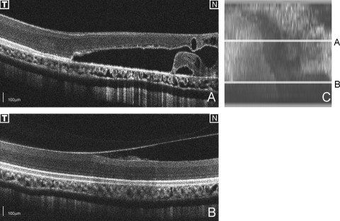
7. What Are Some Examples of Traumatic Injuries to Avian Retinas That Can Be Imaged with SD-OCT?
SD-OCT is invaluable for imaging and diagnosing traumatic injuries to avian retinas. Examples of such injuries include:
- Retinal Detachment (RD): SD-OCT can clearly visualize retinal detachment, including the separation of the retina from the retinal pigment epithelium (RPE) and the presence of subretinal fluid.
- Retinal Lesions: SD-OCT can identify lesions, thinning, and atrophy in the retina, providing detailed information about the extent and location of the damage.
- Intraretinal Cysts: SD-OCT can detect intraretinal cystic spaces, which may form as a result of retinal degeneration or injury.
- Choroidal Damage: SD-OCT can reveal damage to the choroid, the vascular layer beneath the retina, which can affect retinal health.
- Pecten Abnormalities: SD-OCT can image abnormalities in the pecten oculi, such as inflammation or degeneration, which can impair its function.
8. How Does the Distribution of the Retinal Nerve Fiber Layer (RNFL) Relate to the Fovea in Birds?
The distribution of the retinal nerve fiber layer (RNFL) is closely related to the fovea in birds. The RNFL is composed of axons from ganglion cells, which transmit visual information from the retina to the brain. Key features include:
- RNFL Avoidance: The RNFL typically avoids the foveal region to minimize light scattering and maximize visual acuity. This avoidance creates a characteristic thinning of the RNFL around the fovea.
- Perifoveal Thickening: In some species, there may be a thickening of the RNFL in the perifoveal region, which is thought to enhance signal processing and visual sensitivity.
- RNFL Thickness Mapping: Mapping RNFL thickness can help researchers identify the location and extent of the fovea and understand its relationship to the optic nerve head (ONH).
- Composite Imaging: Combining multiple datasets allows for the creation of composite RNFL thickness maps, providing a comprehensive view of the RNFL distribution across the retina.
9. What Variations Exist in the Topography of the Retina Among Different Species of Raptors?
Significant variations exist in the topography of the retina among different species of raptors, reflecting their diverse hunting strategies and visual adaptations:
- Foveal Morphology: As noted earlier, the depth, shape, and location of the fovea can vary considerably. Some raptors have deep, steep foveas for high-resolution vision, while others have shallower foveas for enhanced motion detection.
- Retinal Thickness: The overall thickness of the retina and individual retinal layers can vary, influencing visual sensitivity and acuity.
- Presence of Multiple Foveas: Some raptors have multiple foveas, providing a wider field of high-acuity vision and enhancing their ability to track prey.
- Choroidal Structure: The structure and thickness of the choroid, the vascular layer beneath the retina, can also vary, affecting retinal health and oxygen supply.
- Pecten Size and Shape: The size and shape of the pecten oculi can differ, influencing its effectiveness in nourishing the retina and reducing shadows.
10. What Clinical Applications Might Arise From Vision Research in Birds of Prey?
Vision research in birds of prey has potential clinical applications for both avian and human ophthalmology:
- Understanding Visual Acuity: Studying the mechanisms that enable the exceptional visual acuity of birds of prey can provide insights into improving vision in humans with visual impairments.
- Retinal Disease Treatment: Research on avian retinal structures and diseases can lead to new treatments for human retinal disorders, such as macular degeneration and retinal detachment.
- Diagnostic Techniques: Techniques developed for imaging avian retinas, such as SD-OCT, can be adapted for use in human ophthalmology, improving the diagnosis and management of eye diseases.
- Comparative Anatomy: Understanding the comparative anatomy of avian and human eyes can provide insights into the evolution and function of the visual system.
- Conservation Efforts: Vision research can aid in the conservation of endangered bird species by providing information about their visual needs and the impact of environmental factors on their vision.
11. Why Do Some Birds Have a Thicker Photoreceptor Outer Segment?
A thicker photoreceptor outer segment (OS) in some birds typically indicates a higher concentration of photopigments, enhancing light sensitivity. This adaptation is particularly common in nocturnal birds, where increased light sensitivity is crucial for vision in low-light conditions. The outer segment contains the light-sensitive molecules (rhodopsin in rods and cone pigments in cones) that initiate the visual process. A thicker OS means more of these molecules are present, allowing the bird to capture more photons and see better in dim environments.
12. How Does the Ganglion Cell Layer Contribute to Vision in Avian Species?
The ganglion cell layer (GCL) is the innermost layer of the retina containing ganglion cells, which are responsible for transmitting visual information from the retina to the brain via the optic nerve. In avian species, the GCL plays a crucial role in:
- Signal Processing: Ganglion cells process and integrate signals from photoreceptors and other retinal neurons, enhancing contrast and detecting motion.
- Spatial Resolution: The density and arrangement of ganglion cells influence spatial resolution and visual acuity.
- Color Vision: Different types of ganglion cells are involved in processing color information, contributing to the vibrant color vision of many birds.
- Circadian Rhythms: Some ganglion cells contain melanopsin, a photopigment that is sensitive to ambient light and helps regulate circadian rhythms and other non-visual functions.
Variations in the GCL structure and function can reflect adaptations to different visual tasks and ecological niches.
13. What Are the Key Differences in Retinal Vascularization Between Birds and Mammals?
A significant difference between the eyes of birds and mammals is their retinal vascularization. Mammals have a vascularized retina, meaning that blood vessels run across the surface of the retina to supply it with oxygen and nutrients. In contrast, birds have an avascular retina, lacking blood vessels on the retinal surface. Instead, birds rely on two primary mechanisms for retinal nourishment and oxygenation:
- Pecten Oculi: As mentioned earlier, the pecten oculi provides nourishment and oxygen to the retina.
- Choriocapillaris: Oxygenation is received from the choriocapillaris, a dense network of blood vessels in the choroid layer beneath the retina.
The avascular nature of the avian retina minimizes light scattering, enhancing visual clarity and acuity, which is essential for their visual tasks.
14. How Can RNFL Thickness Maps Aid in Diagnosing Glaucoma in Birds?
RNFL thickness maps can be valuable tools for diagnosing glaucoma in birds, similar to their use in human ophthalmology. Glaucoma is a condition characterized by damage to the optic nerve, often associated with increased intraocular pressure. Key applications include:
- Detecting RNFL Thinning: Glaucoma typically causes thinning of the RNFL as ganglion cell axons are damaged. RNFL thickness maps can reveal localized or diffuse areas of thinning, indicating glaucomatous damage.
- Monitoring Disease Progression: Serial RNFL thickness maps can be used to monitor the progression of glaucoma over time, assessing the effectiveness of treatment interventions.
- Differentiating Glaucoma from Other Conditions: RNFL thickness maps can help differentiate glaucoma from other conditions that may cause optic nerve damage, such as optic neuritis or retinal ischemia.
- Early Detection: RNFL thickness maps can detect subtle changes in the RNFL before significant visual field loss occurs, allowing for early intervention and treatment.
However, it’s important to note that the interpretation of RNFL thickness maps in birds requires careful consideration of species-specific anatomical variations and the establishment of normative data.
15. How Does the Location of the Fovea Affect a Bird’s Visual Field?
The location of the fovea on the retina significantly affects a bird’s visual field and how it perceives its surroundings. Variations in foveal location include:
- Central Fovea: A central fovea provides high-acuity vision in the center of the visual field, ideal for tasks requiring detailed observation directly ahead.
- Temporal Fovea: A temporal fovea enhances peripheral vision, allowing birds to detect movement and potential threats from the sides. This is particularly useful for birds that need to maintain awareness of their surroundings while foraging or flying.
- Nasal Fovea: A nasal fovea is positioned towards the midline of the head, providing enhanced binocular vision and depth perception in the forward field of view.
- Multiple Foveas: Species with multiple foveas can combine the advantages of different foveal locations, providing a wide field of high-acuity vision and enhanced depth perception.
The optimal foveal location depends on the bird’s lifestyle, hunting strategy, and ecological niche.
16. What Environmental Factors Can Influence the Development and Structure of Avian Foveas?
Several environmental factors can influence the development and structure of avian foveas:
- Light Availability: The amount and quality of light available in a bird’s habitat can affect the development of photoreceptors and the overall structure of the fovea.
- Diet: Nutritional factors, such as the availability of certain vitamins and antioxidants, can influence retinal health and foveal development.
- Visual Tasks: The types of visual tasks a bird performs (e.g., hunting, foraging, navigating) can drive the evolution and adaptation of its foveal structure.
- Predation Pressure: The risk of predation can influence the development of foveas optimized for motion detection and peripheral vision.
- Habitat Complexity: Birds living in complex habitats may develop foveas adapted for detailed spatial vision and depth perception.
These environmental factors can exert selective pressures that shape the evolution of avian foveas, optimizing visual performance for survival and reproduction.
17. How Does the Optical Design of the Avian Eye Contribute to High Visual Acuity?
The optical design of the avian eye is finely tuned to contribute to high visual acuity. Key features include:
- Avascular Retina: The absence of blood vessels on the retinal surface minimizes light scattering, enhancing visual clarity.
- Pecten Oculi: The pecten oculi reduces shadows cast by blood vessels and provides retinal nourishment.
- Lens Shape: The shape and refractive properties of the lens optimize light focusing on the fovea.
- Corneal Curvature: The curvature of the cornea contributes to light refraction and image formation.
- Eye Size: Larger eyes generally have better spatial resolution, allowing for finer detail detection.
These optical adaptations work in concert to maximize light capture, minimize distortion, and focus a sharp image on the retina, contributing to the exceptional visual acuity of many birds.
18. What Are Some Ethical Considerations in Vision Research Involving Birds of Prey?
Ethical considerations are paramount in vision research involving birds of prey. Key considerations include:
- Animal Welfare: Ensuring the welfare of the birds is essential. This includes providing appropriate housing, nutrition, and veterinary care.
- Minimizing Stress: Research protocols should be designed to minimize stress and discomfort to the birds.
- Non-Invasive Techniques: Prioritize non-invasive imaging techniques, such as SD-OCT, to minimize harm to the birds.
- IACUC Approval: All research protocols should be reviewed and approved by an Institutional Animal Care and Use Committee (IACUC) to ensure compliance with ethical guidelines and regulations.
- Justification of Research: The scientific value and potential benefits of the research should justify the use of birds of prey.
- Conservation Impact: Research should consider the potential impact on the conservation of endangered species and contribute to their protection.
Adhering to these ethical considerations is crucial for conducting responsible and humane vision research involving birds of prey.
19. Can Avian Vision Research Help in Developing Better Artificial Vision Systems for Humans?
Yes, avian vision research can provide valuable insights for developing better artificial vision systems for humans. By studying the unique adaptations of the avian visual system, researchers can gain knowledge that can be applied to the design of artificial retinas and other vision prosthetics. Specific contributions include:
- Photoreceptor Design: Understanding the structure and function of avian photoreceptors can inspire the design of more efficient and sensitive artificial photoreceptors.
- Signal Processing Algorithms: Studying how the avian retina processes visual information can inform the development of more sophisticated signal processing algorithms for artificial vision systems.
- Avascular Retina Models: The avascular nature of the avian retina can provide insights into how to design artificial retinas that do not rely on blood vessels for nourishment, minimizing light scattering and improving image quality.
- Foveal Mimicry: Research on avian foveas can guide the design of artificial retinas with enhanced spatial resolution in the central visual field.
- Motion Detection: Studying how birds detect motion can lead to the development of artificial vision systems with improved motion detection capabilities.
20. What Are the Future Directions in Avian Fovea Research?
Future directions in avian fovea research include:
- Advanced Imaging Techniques: Utilizing advanced imaging techniques, such as adaptive optics OCT, to visualize foveal structures at even higher resolution.
- Genetic Studies: Investigating the genetic basis of foveal development and adaptation in different bird species.
- Behavioral Studies: Combining anatomical and physiological data with behavioral studies to better understand how foveal structure relates to visual performance in natural environments.
- Comparative Studies: Conducting more comprehensive comparative studies across a wider range of bird species to understand the diversity of foveal adaptations.
- Conservation Applications: Applying fovea research to conservation efforts by assessing the impact of environmental factors on avian vision and identifying threats to visual health.
These future research directions promise to provide a deeper understanding of the avian fovea and its role in vision, with implications for both basic science and applied conservation efforts.
At COMPARE.EDU.VN, we understand the challenges in comparing complex information. Our goal is to provide you with clear, detailed comparisons to help you make informed decisions. Explore our site to find more comparisons and resources.
Contact us:
Address: 333 Comparison Plaza, Choice City, CA 90210, United States
Whatsapp: +1 (626) 555-9090
Website: compare.edu.vn
FAQ: Deep Avian Foveas
- What is the main purpose of a deep avian fovea?
The primary function is to provide high-acuity vision, crucial for tasks like hunting and detailed observation. - How does the deep avian fovea differ from the shallow avian fovea?
The deep avian fovea is used for high-resolution, binocular vision, while the shallow avian fovea is mainly for monocular vision and motion detection. - Why do diurnal birds have more cones in their foveas compared to nocturnal birds?
Diurnal birds require excellent color vision and high visual acuity in bright light, hence the higher cone density. - What role does the pecten oculi play in the avian eye?
The pecten provides nourishment and oxygen to the retina, reduces shadows, and helps regulate eye pressure. - How does SD-OCT enhance the study of avian retinal structures?
SD-OCT provides high-resolution, non-invasive imaging of the retina, allowing detailed visualization of retinal layers and structures. - What kind of traumatic injuries can be imaged with SD-OCT in avian retinas?
Retinal detachment, lesions, intraretinal cysts, and choroidal damage can be effectively imaged with SD-OCT. - How is the retinal nerve fiber layer (RNFL) related to the fovea?
The RNFL typically avoids the foveal region to minimize light scattering and maximize visual acuity. - What are some ethical considerations when conducting vision research on birds of prey?
Ensuring animal welfare, minimizing stress, using non-invasive techniques, and justifying the research are key ethical considerations. - Can avian vision research help in developing better artificial vision systems for humans?
Yes, insights from avian vision research can inform the design of more efficient and sensitive artificial photoreceptors and signal processing algorithms. - What are some future directions in avian fovea research?
Future research includes utilizing advanced imaging techniques, genetic studies, and behavioral studies to understand the diversity of foveal adaptations.