In the anatomical position, the fingers are distal to the elbow, meaning they are further away from the trunk of the body. This positioning is crucial for healthcare professionals and anyone studying anatomy. At COMPARE.EDU.VN, we delve into the intricacies of anatomical terminology and positional relationships. Understanding anatomical terms and spatial relationships helps prevent medical errors and ensure accurate communications.
1. Understanding Anatomical Position
Anatomical position serves as the universal reference point in anatomy and medicine. It ensures consistency and clarity in describing the location of body parts and structures.
1.1 Defining Anatomical Position
The anatomical position is a standardized reference posture. The body is upright, feet are parallel and flat on the floor, eyes are forward, and the upper limbs are at the sides with palms facing forward.
1.2 Significance of Anatomical Position
The anatomical position is vital for:
- Accurate Communication: Healthcare professionals use it to precisely describe the location of injuries, abnormalities, or structures, minimizing ambiguity.
- Standardized Reference: Provides a common frame of reference in anatomical studies, research, and clinical practice.
- Consistency: Ensures that descriptions are universally understood, regardless of individual variations in posture.
2. Anatomical Terminology: Directional Terms
Directional terms are essential for accurately describing the location of one body part relative to another. These terms are always based on the anatomical position, providing a consistent frame of reference.
2.1 Key Directional Terms
- Superior (Cranial): Closer to the head.
- Inferior (Caudal): Closer to the feet.
- Anterior (Ventral): Towards the front of the body.
- Posterior (Dorsal): Towards the back of the body.
- Medial: Closer to the midline of the body.
- Lateral: Further from the midline of the body.
- Proximal: Closer to the point of attachment to the trunk.
- Distal: Further from the point of attachment to the trunk.
- Superficial: Closer to the surface of the body.
- Deep: Further from the surface of the body.
2.2 Applying Directional Terms
Understanding how to apply directional terms is crucial for clear anatomical description. For example:
- The nose is superior to the mouth.
- The stomach is anterior to the spine.
- The thumb is lateral to the index finger.
- The shoulder is proximal to the elbow.
3. The Upper Limb: Elbow and Fingers
The upper limb consists of several segments, including the arm, forearm, and hand. Understanding the relationship between these segments is essential for describing movements and positions accurately.
3.1 Anatomy of the Elbow
The elbow is a hinge joint that connects the upper arm (humerus) to the forearm (radius and ulna). It allows for flexion and extension movements. Key structures include:
- Humerus: The bone of the upper arm.
- Radius: One of the two bones of the forearm, located on the thumb side.
- Ulna: The other bone of the forearm, located on the pinky side.
- Elbow Joint: The articulation between the humerus, radius, and ulna.
3.2 Anatomy of the Fingers
The fingers are located at the distal end of the upper limb and are part of the hand. Each finger consists of three phalanges (except for the thumb, which has two). Key structures include:
- Metacarpals: The bones of the hand that articulate with the phalanges.
- Phalanges: The bones of the fingers (proximal, middle, and distal).
- Interphalangeal Joints: The joints between the phalanges, allowing for flexion and extension.
- Metacarpophalangeal Joints: The joints between the metacarpals and the proximal phalanges, allowing for a wider range of movements.
4. Positional Relationship: Fingers and Elbow
To accurately describe the positional relationship between the fingers and the elbow, we use the directional term “distal.”
4.1 Distal Definition and Application
Distal means “further from the point of attachment to the trunk.” In the context of the upper limb, the fingers are located further away from the trunk compared to the elbow.
4.2 Fingers are Distal to the Elbow
When the body is in the anatomical position, the fingers are distal to the elbow. This means that the fingers are further down the limb, away from the shoulder (the point of attachment to the trunk), than the elbow.
4.3 Implications of This Relationship
Understanding this distal relationship is critical in various contexts:
- Medical Diagnosis: If a patient reports pain radiating from the elbow down to the fingers, it helps healthcare providers understand potential nerve or vascular involvement.
- Physical Therapy: Rehabilitation exercises often focus on restoring proper function from proximal to distal regions, or vice versa, to improve overall limb function.
- Ergonomics: Understanding the reach and movement capabilities of the hand relative to the elbow is essential in designing workspaces and tools.
5. Other Relevant Anatomical Relationships in the Upper Limb
Understanding additional anatomical relationships can provide a more comprehensive understanding of the upper limb.
5.1 Proximal and Distal Relationships
- Shoulder vs. Elbow: The shoulder is proximal to the elbow.
- Wrist vs. Fingers: The wrist is proximal to the fingers.
- Elbow vs. Wrist: The elbow is proximal to the wrist.
5.2 Superior and Inferior Relationships
- Shoulder vs. Wrist: The shoulder is superior to the wrist.
- Elbow vs. Fingers: The elbow is superior to the fingers.
5.3 Medial and Lateral Relationships
- Ulna vs. Radius: The ulna is medial to the radius in the anatomical position.
- Thumb vs. Pinky: The thumb is lateral to the pinky finger in the anatomical position.
6. Movements of the Upper Limb
Understanding the movements possible at the joints of the upper limb further clarifies the relationships between its parts.
6.1 Movements at the Elbow Joint
The elbow joint primarily allows for flexion and extension:
- Flexion: Decreasing the angle between the forearm and the upper arm.
- Extension: Increasing the angle between the forearm and the upper arm, straightening the elbow.
6.2 Movements at the Wrist Joint
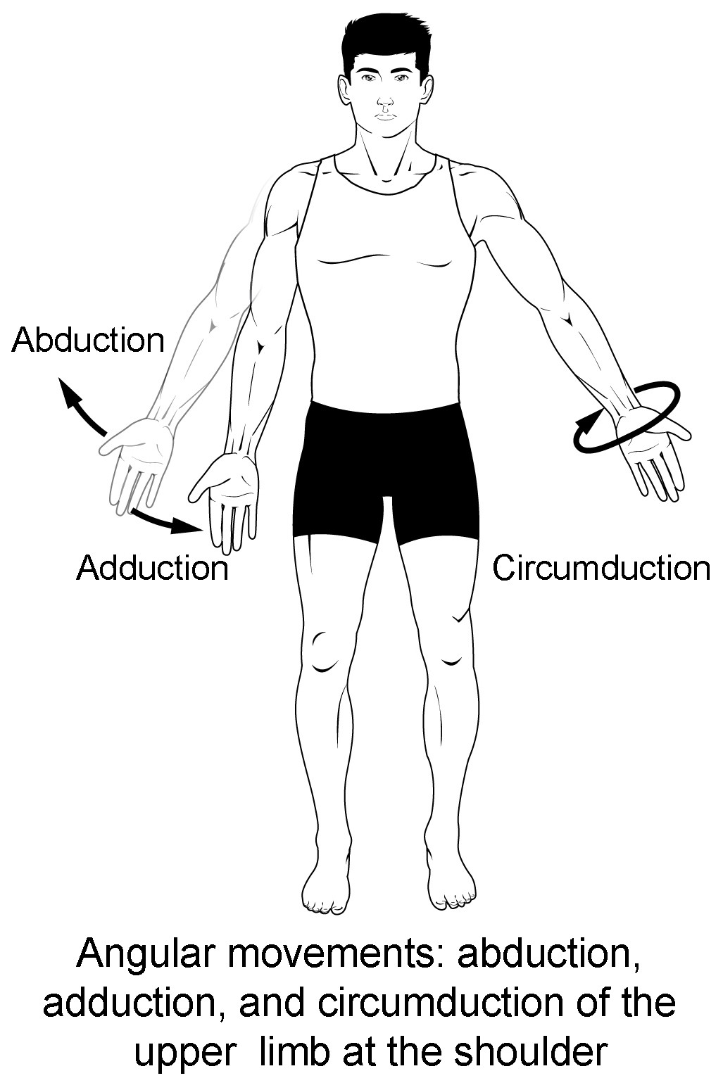
The wrist joint allows for flexion, extension, abduction (radial deviation), and adduction (ulnar deviation):
- Flexion: Bending the hand towards the palm.
- Extension: Bending the hand towards the back of the hand.
- Abduction (Radial Deviation): Moving the hand laterally, towards the thumb.
- Adduction (Ulnar Deviation): Moving the hand medially, towards the pinky.
6.3 Movements at the Finger Joints
The finger joints (metacarpophalangeal and interphalangeal) allow for flexion, extension, abduction, and adduction:
- Flexion: Bending the fingers towards the palm.
- Extension: Straightening the fingers.
- Abduction: Spreading the fingers apart from the midline of the hand.
- Adduction: Bringing the fingers together towards the midline of the hand.
7. Clinical Significance of Anatomical Relationships
The anatomical relationships between body parts are not just theoretical concepts; they have practical applications in clinical medicine.
7.1 Nerve and Vascular Pathways
Nerves and blood vessels follow specific pathways in the body. Understanding these pathways and their relationship to anatomical structures is crucial for diagnosing and treating nerve injuries or vascular disorders.
- Median Nerve: The median nerve passes through the elbow and extends down into the forearm and hand. Compression of this nerve at the wrist can lead to carpal tunnel syndrome, affecting the function of the thumb and first two fingers.
- Ulnar Nerve: The ulnar nerve passes behind the medial epicondyle of the humerus at the elbow. Compression or irritation of this nerve can cause cubital tunnel syndrome, affecting the pinky and ring fingers.
- Radial Artery: The radial artery travels down the lateral side of the forearm and provides blood supply to the hand. Its pulse can be palpated at the wrist, distal to the elbow.
7.2 Musculoskeletal Injuries
Understanding the anatomical relationships helps in diagnosing and treating musculoskeletal injuries.
- Elbow Dislocation: A dislocation at the elbow joint can affect the position and function of the forearm and hand, potentially impacting the fingers.
- Fractures: Fractures of the humerus, radius, or ulna can alter the anatomical relationships between the elbow and the fingers, requiring precise realignment during treatment.
- Tendonitis: Inflammation of the tendons around the elbow (e.g., tennis elbow or golfer’s elbow) can cause pain that radiates down the forearm towards the wrist and fingers.
8. Common Anatomical Variations
While the anatomical position provides a standard reference, it’s important to recognize that anatomical variations exist among individuals.
8.1 Variations in Muscle Attachments
The precise attachment points of muscles can vary, affecting the range of motion and strength at joints like the elbow and wrist.
8.2 Variations in Nerve Pathways
Nerve pathways can also vary. For example, the course of the median or ulnar nerve may differ slightly in some individuals, which can affect the symptoms of nerve compression syndromes.
8.3 Importance of Recognizing Variations
Healthcare professionals must be aware of these potential variations to accurately diagnose and treat patients. Imaging techniques like MRI and ultrasound can help identify anatomical variations before surgical interventions.
9. Anatomical Planes
Anatomical planes are imaginary flat surfaces that divide the body, providing additional reference points for describing the location and direction of structures and movements.
9.1 Sagittal Plane
The sagittal plane divides the body into right and left portions. Movements in this plane include flexion and extension.
9.2 Frontal (Coronal) Plane
The frontal plane divides the body into anterior and posterior portions. Movements in this plane include abduction and adduction.
9.3 Transverse (Axial) Plane
The transverse plane divides the body into superior and inferior portions. Movements in this plane include rotation.
9.4 Using Planes to Describe Movements
Understanding anatomical planes helps in describing movements accurately. For example:
- Flexion and extension of the elbow occur in the sagittal plane.
- Abduction and adduction of the fingers occur in the frontal plane.
- Pronation and supination of the forearm involve rotation in the transverse plane.
10. Additional Terms of Movement
Beyond the basic movements, some specific terms describe more complex actions at joints.
10.1 Circumduction
Circumduction is a circular movement that combines flexion, abduction, extension, and adduction. It is possible at the shoulder, hip, wrist, and metacarpophalangeal joints.
10.2 Rotation
Rotation involves turning a bone around its longitudinal axis. Medial (internal) rotation turns the anterior surface towards the midline, while lateral (external) rotation turns it away.
10.3 Pronation and Supination
Pronation and supination are specific to the forearm. Pronation turns the palm backward, while supination turns it forward.
10.4 Inversion and Eversion
Inversion and eversion are movements of the foot at the intertarsal joints. Inversion turns the sole of the foot inward, while eversion turns it outward.
10.5 Protraction and Retraction
Protraction and retraction are anterior and posterior movements in the transverse plane. Protraction moves a structure forward (e.g., protracting the jaw), while retraction moves it backward.
10.6 Elevation and Depression
Elevation and depression involve moving a structure superiorly (elevation) or inferiorly (depression). These movements are commonly used to describe movements of the scapula or mandible.
11. Comprehensive Review of Anatomical Terminology
Reinforcing the key anatomical terms can improve retention and application in various contexts.
11.1 Summary of Directional Terms
- Superior: Towards the head.
- Inferior: Towards the feet.
- Anterior: Towards the front.
- Posterior: Towards the back.
- Medial: Towards the midline.
- Lateral: Away from the midline.
- Proximal: Closer to the trunk.
- Distal: Further from the trunk.
- Superficial: Closer to the surface.
- Deep: Further from the surface.
11.2 Summary of Movement Terms
- Flexion: Decreasing the angle at a joint.
- Extension: Increasing the angle at a joint.
- Abduction: Moving away from the midline.
- Adduction: Moving towards the midline.
- Rotation: Turning around the longitudinal axis.
- Circumduction: Circular movement combining flexion, abduction, extension, and adduction.
- Pronation: Turning the palm backward.
- Supination: Turning the palm forward.
- Inversion: Turning the sole of the foot inward.
- Eversion: Turning the sole of the foot outward.
- Protraction: Moving forward.
- Retraction: Moving backward.
- Elevation: Moving superiorly.
- Depression: Moving inferiorly.
11.3 Application in Describing Clinical Scenarios
Consider the following clinical scenarios and how anatomical terminology applies:
- Scenario 1: A patient reports pain on the lateral side of the elbow that radiates distally towards the wrist. This suggests possible lateral epicondylitis (tennis elbow) with referred pain.
- Scenario 2: A patient has difficulty abducting and adducting their fingers. This could indicate a problem with the ulnar nerve, which controls the intrinsic muscles of the hand responsible for these movements.
- Scenario 3: A patient has a fracture of the distal radius. This injury is located proximal to the wrist and distal to the elbow, affecting the forearm.
12. Advancements in Anatomical Studies
Modern technologies have significantly advanced the study and understanding of anatomy.
12.1 Imaging Techniques
- MRI (Magnetic Resonance Imaging): Provides detailed images of soft tissues, including muscles, ligaments, and nerves.
- CT (Computed Tomography) Scan: Provides cross-sectional images of bones and soft tissues.
- Ultrasound: Uses sound waves to create real-time images of anatomical structures.
- X-rays: Provide images of bones and can help identify fractures or dislocations.
12.2 3D Modeling and Virtual Reality
3D modeling and virtual reality (VR) technologies are transforming anatomical education. They allow students and healthcare professionals to explore anatomical structures in an interactive and immersive environment.
12.3 Anatomical Software and Resources
Various software programs and online resources provide detailed anatomical information, including 3D models, diagrams, and animations. These tools enhance the learning experience and make it easier to visualize complex anatomical relationships.
13. The Role of Anatomy in Different Professions
A strong understanding of anatomy is crucial in various professions.
13.1 Medical Doctors and Surgeons
Medical doctors and surgeons rely on anatomical knowledge to diagnose and treat diseases, perform surgeries, and administer medications. Accurate anatomical knowledge is essential for avoiding iatrogenic injuries during medical procedures.
13.2 Physical Therapists and Occupational Therapists
Physical therapists and occupational therapists use anatomical knowledge to assess and treat musculoskeletal injuries, restore function, and improve patients’ quality of life. They design exercise programs based on anatomical principles to target specific muscles and joints.
13.3 Athletic Trainers
Athletic trainers use anatomical knowledge to prevent and treat sports-related injuries. They understand the biomechanics of movement and can identify potential risk factors for injuries based on anatomical variations.
13.4 Massage Therapists
Massage therapists use anatomical knowledge to target specific muscles and soft tissues to relieve pain, reduce muscle tension, and improve circulation. They understand the location and function of muscles and can apply massage techniques to address specific musculoskeletal problems.
14. Ergonomics and Anatomy
Ergonomics, the study of designing workplaces and equipment to fit the human body, relies heavily on anatomical principles.
14.1 Importance of Ergonomic Design
Proper ergonomic design can reduce the risk of musculoskeletal disorders, improve productivity, and enhance comfort.
14.2 Ergonomic Considerations for the Upper Limb
When designing workstations and tools, it’s important to consider the anatomical relationships between the shoulder, elbow, wrist, and fingers.
- Elbow Angle: Maintaining a neutral elbow angle (around 90 degrees) can reduce strain on the elbow joint and surrounding muscles.
- Wrist Position: Keeping the wrist in a neutral position can prevent carpal tunnel syndrome and other wrist injuries.
- Finger Reach: Ensuring that frequently used items are within easy reach can reduce strain on the fingers and hand.
14.3 Examples of Ergonomic Interventions
- Adjustable Chairs: Allow users to adjust the height and back support to maintain proper posture.
- Keyboard Trays: Position the keyboard at a comfortable height and angle to reduce wrist strain.
- Ergonomic Mice: Designed to fit the natural shape of the hand and reduce strain on the wrist and fingers.
15. Future Trends in Anatomical Education
Anatomical education is continuously evolving with advancements in technology and changes in healthcare practices.
15.1 Virtual Dissection Tables
Virtual dissection tables use 3D imaging technology to simulate the experience of dissecting a human cadaver without the need for real cadavers. These tables allow students to explore anatomical structures in a realistic and interactive way.
15.2 Augmented Reality (AR) Applications
Augmented reality (AR) applications overlay digital information onto the real world, allowing students to visualize anatomical structures on their own bodies or on anatomical models.
15.3 Online Anatomical Resources
Online anatomical resources, including videos, interactive modules, and virtual labs, provide students with access to anatomical information anytime, anywhere. These resources enhance the learning experience and make it easier to review and reinforce anatomical concepts.
16. Maintaining Proper Posture for Optimal Anatomy
Good posture is essential for maintaining proper anatomical alignment and preventing musculoskeletal problems.
16.1 Importance of Good Posture
Good posture helps distribute weight evenly across the body, reducing strain on muscles and joints.
16.2 Tips for Maintaining Good Posture
- Sit Upright: When sitting, keep your back straight, shoulders relaxed, and feet flat on the floor.
- Stand Tall: When standing, keep your head level, shoulders relaxed, and weight evenly distributed on both feet.
- Take Breaks: Take frequent breaks to stretch and move around, especially if you spend long periods sitting or standing in the same position.
- Exercise Regularly: Regular exercise can strengthen the muscles that support good posture.
16.3 Common Postural Problems
- Forward Head Posture: Occurs when the head is positioned forward of the shoulders, leading to neck pain and stiffness.
- Rounded Shoulders: Occur when the shoulders are rolled forward, leading to upper back pain and limited range of motion.
- Swayback Posture: Occurs when the pelvis is tilted forward, leading to lower back pain and muscle imbalances.
17. Anatomical Terminology and Imaging Modalities
Understanding how anatomical structures appear on different imaging modalities is essential for accurate diagnosis and treatment.
17.1 Anatomical Landmarks on X-Rays
X-rays are commonly used to visualize bones. Key anatomical landmarks on upper limb X-rays include:
- Humerus: The bone of the upper arm, extending from the shoulder to the elbow.
- Radius: The lateral bone of the forearm, extending from the elbow to the wrist.
- Ulna: The medial bone of the forearm, extending from the elbow to the wrist.
- Carpals: The bones of the wrist, located between the forearm and the hand.
- Metacarpals: The bones of the hand, extending from the wrist to the fingers.
- Phalanges: The bones of the fingers.
17.2 Anatomical Structures on MRI
MRI provides detailed images of soft tissues. Key anatomical structures on upper limb MRIs include:
- Muscles: The muscles of the upper arm, forearm, and hand.
- Tendons: The tendons that attach muscles to bones.
- Ligaments: The ligaments that stabilize the joints.
- Nerves: The nerves that innervate the upper limb, including the median, ulnar, and radial nerves.
- Blood Vessels: The arteries and veins that supply blood to the upper limb.
17.3 Anatomical Visualization on Ultrasound
Ultrasound uses sound waves to create real-time images of anatomical structures. It is commonly used to visualize tendons, ligaments, and nerves.
18. Biomechanics of Upper Limb Movement
Understanding the biomechanics of upper limb movement involves studying the forces and mechanics that control movement at the joints.
18.1 Lever Systems in the Upper Limb
The upper limb utilizes lever systems to generate force and movement. Lever systems consist of a fulcrum (joint), a force (muscle contraction), and a load (resistance).
18.2 Muscle Actions at the Elbow Joint
- Flexion: The primary flexors of the elbow joint are the biceps brachii, brachialis, and brachioradialis muscles.
- Extension: The primary extensor of the elbow joint is the triceps brachii muscle.
- Supination: The primary supinator of the forearm is the biceps brachii muscle.
- Pronation: The primary pronators of the forearm are the pronator teres and pronator quadratus muscles.
18.3 Muscle Actions at the Wrist and Finger Joints
- Wrist Flexion: The primary wrist flexors are the flexor carpi radialis and flexor carpi ulnaris muscles.
- Wrist Extension: The primary wrist extensors are the extensor carpi radialis longus, extensor carpi radialis brevis, and extensor carpi ulnaris muscles.
- Finger Flexion: The primary finger flexors are the flexor digitorum superficialis and flexor digitorum profundus muscles.
- Finger Extension: The primary finger extensors are the extensor digitorum, extensor indicis, and extensor digiti minimi muscles.
19. Common Pathologies of the Upper Limb
Understanding common pathologies of the upper limb requires knowledge of anatomy, biomechanics, and clinical medicine.
19.1 Carpal Tunnel Syndrome
Carpal tunnel syndrome is a common condition caused by compression of the median nerve at the wrist. Symptoms include pain, numbness, and tingling in the thumb, index finger, and middle finger.
19.2 Cubital Tunnel Syndrome
Cubital tunnel syndrome is caused by compression of the ulnar nerve at the elbow. Symptoms include pain, numbness, and tingling in the pinky and ring finger.
19.3 Lateral Epicondylitis (Tennis Elbow)
Lateral epicondylitis is an inflammation of the tendons on the lateral side of the elbow. Symptoms include pain and tenderness on the outer side of the elbow.
19.4 Medial Epicondylitis (Golfer’s Elbow)
Medial epicondylitis is an inflammation of the tendons on the medial side of the elbow. Symptoms include pain and tenderness on the inner side of the elbow.
19.5 De Quervain’s Tenosynovitis
De Quervain’s tenosynovitis is a condition that affects the tendons on the thumb side of the wrist. Symptoms include pain and tenderness at the base of the thumb.
20. The Importance of Anatomical Terminology in Patient Care
Clear and accurate communication using anatomical terminology is essential for providing safe and effective patient care.
20.1 Preventing Medical Errors
Using anatomical terminology helps prevent misunderstandings and errors in communication between healthcare professionals.
20.2 Ensuring Accurate Documentation
Accurate documentation using anatomical terminology ensures that patient records are clear, concise, and informative.
20.3 Enhancing Patient Understanding
Explaining anatomical concepts to patients using simple and understandable language can help them better understand their conditions and treatment plans.
20.4 Fostering Interprofessional Communication
Using anatomical terminology promotes effective communication between different healthcare professionals, such as doctors, nurses, therapists, and athletic trainers.
Understanding the anatomical relationships between the fingers and elbow is fundamental in medicine, therapy, and ergonomics. Remember, the fingers are distal to the elbow in the anatomical position. At COMPARE.EDU.VN, we strive to provide comprehensive and accessible information to help you make informed decisions and expand your knowledge. For more detailed comparisons and resources, visit our website at COMPARE.EDU.VN or contact us at 333 Comparison Plaza, Choice City, CA 90210, United States or Whatsapp: +1 (626) 555-9090. Explore anatomical placements, directional terminology, and musculoskeletal system details for a deeper understanding of the human body.
 A diagram showing anatomical positions, including anterior, posterior, medial, lateral, proximal, and distal.
FAQ: Anatomical Position of Fingers Compared to Elbow
1. In the anatomical position, what does “distal” mean?
Distal refers to being farther away from the point of attachment to the trunk of the body.
2. How does the anatomical position relate to describing body parts?
The anatomical position provides a standardized reference point for accurately describing the location and relationship of body parts.
3. Why is it important to use anatomical terminology correctly?
Correct use of anatomical terminology ensures clear communication, prevents medical errors, and promotes accurate documentation in healthcare settings.
4. Can you give an example of how anatomical position is used in clinical practice?
In describing pain radiating from the elbow down to the fingers, healthcare providers use anatomical terms to understand potential nerve or vascular involvement.
5. How do anatomical planes help in describing body movements?
Anatomical planes (sagittal, frontal, transverse) provide reference points for describing the direction and type of movement at joints.
6. What are some common variations in anatomical structures?
Variations in muscle attachments and nerve pathways are common. Recognizing these variations is important for accurate diagnosis and treatment.
7. What are some imaging techniques used to study anatomical structures?
MRI, CT scans, ultrasound, and X-rays are commonly used to visualize anatomical structures.
8. How does ergonomics utilize anatomical principles?
Ergonomics applies anatomical principles to design workplaces and equipment that fit the human body, reducing the risk of musculoskeletal disorders.
9. What is the role of anatomical knowledge in physical therapy?
Physical therapists use anatomical knowledge to assess and treat musculoskeletal injuries, restore function, and improve patients’ quality of life.
10. How can I learn more about anatomical terminology and relationships?
Visit compare.edu.vn for comprehensive resources, comparisons, and detailed information on anatomical concepts, or contact us at 333 Comparison Plaza, Choice City, CA 90210, United States or Whatsapp: +1 (626) 555-9090.